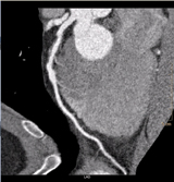
Figure 1(a).
Coronary arteries supply blood to the left ventricular muscle (myocardium). Obstructions within the coronary arteries result in significantly reduced blood flow to the relevant territories of the myocardium, thereby causing damage and impairing its function and eventually resulting in myocardial infarction (MI) or death.
The affected/infarcted territories are typically seen as hypo-dense regions by cardiac computed tomography angiography (cardiac CTA).[1] In the absence of automatic approaches, the assessment of these hypo-dense regions is user-dependent and requires extensive manipulation, with quantification requiring further examination of multiple adjacent slices, making this time-consuming. Hence, there is a need for robust tools that can help with clinical workflow.
The Philips Myocardial Defect Assessment application on IntelliSpace Portal provides visual and quantitative assessment of hypo-dense regions of the myocardium seen from a single gated cardiac CTA scan (retrospectively-gated spiral or Step & Shoot Cardiac). Since this is adjunct information obtained from a single cardiac CTA scan, it does not require any additional scans, thus avoiding any additional increase in radiation dose. Depending on the type of scan performed, the radiation dose could range between 0.6 and 3.5 mSv (if Step & Shoot Cardiac and iterative reconstruction techniques were used) and 8 to 11 mSv (using retrospectively-gated spiral CTA with tube current modulation).[2, 3]
24-Year-Old Male with Chest Pain
Soroka University Medical Center in Beer-Sheva, Israel, has successful experience using the Myocardial Defect Assessment solution. As one example, a 24-year-old male was admitted to the emergency department (ED) for chest pain, with an ECG demonstrating S-T segment elevation in the inferior wall leads. Since the patient reported recent severe flu-like symptoms, the initial diagnosis was myocarditis. Due to his risk factors (smoking and a family history of coronary artery disease [CAD]), cardiac CTA was performed on the Philips Brilliance 64 multi-detector CT (MDCT) to rule out CAD.
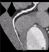
Initial interpretation suggested the presence of a two-vessel disease: There was plaque in the proximal left anterior descending (LAD) (the curved multi-planar reformation is shown in Figure 1a) and also the mid-portion of the right coronary artery (RCA) (the curved multi-planar reformation is shown in Figure 1b). Myocardial Defect Assessment was performed. (An overview of the results is shown in Figure 2.)
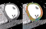
While no hypo-dense regions were observed in the myocardial territories of the LAD, they were present in the basal- and mid-inferior regions of the myocardium (RCA-PLB territories). This is exhibited in the short-axis representation (Figure 3), with the original grayscale images shown on the left, and the corresponding defect probability color representation on the right – the gradient of the color maps represents a range of the myocardial regions that are normal (red) to the hypo-dense regions (blue).
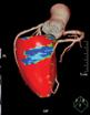
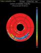
Figure 4(a), rotated to show the RCA-PLB territories, displays the coronary artery tree superimposed on the “egg-shell” color map representation of the LV, with blue indicating hypo-dense regions. Figure 4(b) shows the polar maps representing the short-axis segmentation of LV with the color map overlay using the defect probability method.
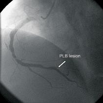
Due to the perfusion finding, a meticulous second look at the PLB territory was performed and a thrombus at the PLB was found. The patient underwent invasive coronary angiography with the lesion area in the mid-RCA stented. Figure 5 shows the focal lesion in the PLB from the invasive coronary angiography.
Case study supplied by Philips Healthcare. Images courtesy of Arik Wolak, M.D., head of cardiac imaging, and Ilan Shelef, M.D., director of Diagnostic Imaging Institute, Soroka University Medical Center.
References:
1. Lessick J, Ghersin E, Dragu R, et al. Diagnostic accuracy of myocardial hypoenhancement on multidetector computed tomography in identifying myocardial infarction in patients admitted with acute chest pain syndrome. J Comput Assist Tomogr. 2007 Sep-Oct; 31(5):780-8.
2. Hou Y, Yue Y, Guo W, et al. Prospectively versus retrospectively ECG-gated 256-slice coronary CT angiography: image quality and radiation dose over expanded heart rates. Int J Cardiovasc Imaging. 2010 Dec 14. [Epub ahead of print]
3. Hosch W, Stiller W, Mueller D, et al. Reduction of radiation exposure and improvement of image quality with BMI-adapted prospective cardiac computed tomography and iterative reconstruction. Eur J Radiol. 2011 Jul 22. [Epub ahead of print]


 April 17, 2024


