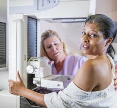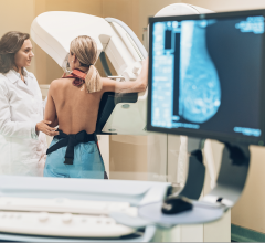
July 1, 2009 - A new breast MRI technique that acquires data radially and generates a high-resolution, three-dimensional image could spare some women with increased breast cancer risk the pain and stress of having to endure a biopsy of a questionable lump or lesion.
The new technique, developed by biomedical engineers at University of Wisconsin-Madison, and part of an ongoing study, could give radiologists greater confidence in visually classifying a lesion as malignant or benign.
To generate the kind of crisp, 3D images necessary for such a diagnosis, Wally Block, a UW-Madison associate professor of biomedical engineering and medical physics, Fred Kelcz, UW-Madison radiology associate professor and graduate student Catherine Moran are capitalizing on their unique MRI data-acquisition method.
An MR image is made up of thousands of smaller pieces of information. The conventional data-acquisition method gathers that information slowly, and it's designed to be viewed from a single imaging plane. Currently, when doing a breast MRI exam, physicians compromise and don't get resolution in the other planes to make it a reasonable scan time. According to Dr. Block, they have found a way around that.
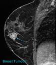
With this new technique, the MRI acquires data radially and generates a high-resolution, three-dimensional image that radiologists can turn, slice and view from many perspectives enabling them to study a lesion's physical characteristics more carefully. Systems equipped with the technique also acquire more data in less time.
In addition, the method also makes it possible for radiologists to view fat images and water images separately, which is particularly useful because fat composes a large portion of the breast.
Block and his colleagues currently are gathering data on the efficacy of the technique. They have tested the method on 20 patients at the University of Wisconsin Hospital and have shared it with colleagues at the University of Toronto for additional assessment. They also are working with Michigan State University researchers to test the technique.
For more information: www.wisc.edu

 February 09, 2024