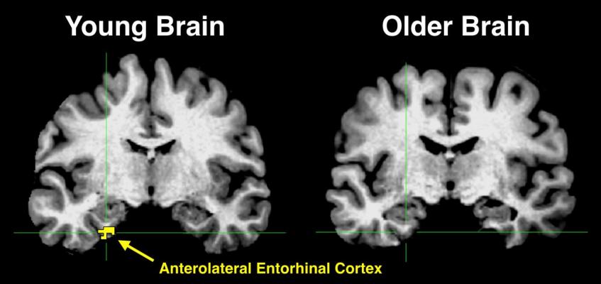
This figure shows two different brains that are aligned to a common template space for comparison. The yellow in the anterolateral entorhinal cortex of the young brain indicates significant activity, something that is absent in the older brain. CREDIT: Zachariah Reagh
As we get older, it's not uncommon to experience "senior moments," in which we forget where we parked our car or call our children by the wrong names. But currently there are no good ways to determine which memory lapses are normal parts of aging and which may signal the early stages of a severe disorder like Alzheimer's disease. In a study appearing March 7 in the journal Neuron, researchers report that data from high-resolution functional brain imaging can be used to show some of the underlying causes for differences in memory proficiency between older and younger adults.
"At the fundamental level, we still understand very little about how aging affects the neural systems that give rise to memory," says Zachariah Reagh, the study's first author, who is now a postdoctoral fellow at the University of California, Davis.
The paper reports data from 20 young adults (ages 18 to 31) and 20 cognitively healthy older adults (ages 64 to 89). The participants were asked to perform two kinds of tasks in an fMRI scanner, an object memory task and a location memory task. Because fMRI looks at the dynamics of blood flow in the brain, it enables investigators to determine which parts of their brains the subjects are using in each task.
In the object task, participants learned pictures of everyday objects and were then asked to distinguish them from new pictures. "Some of the pictures were identical to ones they've seen before, some were brand new, and others were similar to what they've seen before--we may change the color or the size," saids Michael Yassa, Director of the Center for the Neurobiology of Learning and Memory at the University of California, Irvine, and the study's senior author. "We call these tricky items the 'lures.' And we found that older adults struggle with these lures. They are much more likely than younger adults to think they've seen those lures before."
The second task was very similar but required subjects to determine during test whether objects changed their location. For this type of memory task, older adults fared quite a bit better. "This suggests that not all memory changes equally with aging," says Reagh. "Object memory is far more vulnerable than spatial memory, at least in the early stages." Other studies have shown that problems with spatial memory and navigation do manifest as individuals go down the path to Alzheimer's disease.
Importantly, by scanning the subjects' brains while they underwent these tests, the researchers were able to establish a mechanism within the brain for that deficit in object memory.
They found that it was linked to a loss of signaling in the part of the brain called the anterolateral entorhinal cortex. This area was already known to mediate the communication between the hippocampus, where information is first encoded, and the rest of the neocortex, which plays a role in long-term storage. It is also an area that is known to be severely affected in people with Alzheimer's disease.
"The loss of fMRI signal means there is less blood flow to the region, but we believe the underlying basis for this loss has to do with the fact that the structural integrity of that region of the brain is changing," Yassa explained. "One of the things we know about Alzheimer's disease is that this region of the brain is one of the very first to exhibit a key hallmark of the disease, deposition of neurofibrillary tangles."
In contrast, the researchers did not find age-related differences in another area of the brain connected to memory, the posteromedial entorhinal cortex. They demonstrated that this region plays a role in spatial memory, which was also not significantly impaired in the older subjects. "These findings suggest that the brain aging process is selective," Yassa added. "Our findings are not a reflection of general brain aging, but rather specific neural changes that are linked to specific problems in object but not spatial memory."
To determine whether this type of fMRI scan could eventually be used as a tool for early diagnosis, the researchers plan to expand their work to a sample of 150 older adults who will be followed over time. They will also be conducting PET scans to look for amyloid and tau pathology in their brains as they age.
"We hope this comprehensive imaging and cognitive testing will enable us to figure out whether the deficits we saw in the current study are indicative of what is later to come in some of these individuals," Yassa said.
"Our results, as well as similar results from other labs, point to a need for carefully designed tasks and paradigms that can reveal different functions in key areas of the brain and different vulnerabilities to the aging process," Reagh concluded.
For more information: http://www.cell.com/neuron/fulltext/S0896-6273(18)30064-3

 April 17, 2024