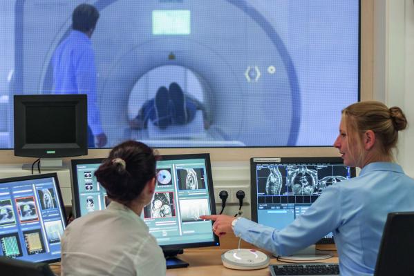
The software developed by Fraunhofer MEVIS automatically identifies the breathing and heart contraction phases in the data independent of the ECG information. This allows for a fast and easy examination of heart patients. Image courtesy of Fraunhofer MEVIS.
November 18, 2015 – Today, magnetic resonance imaging (MRI) allows more gentle, precise and cost-effective heart disease diagnosis. However, the method has limitations when examining children and patients with cardiac arrhythmia. A joint project of the Fraunhofer Institute for Medical Image Computing MEVIS and the Max Planck Institute for Biophysical Chemistry promises a solution for these limitations. Experts were able to accelerate the image acquisition process and expand the method’s applicability.
The importance of MRI for diagnosing cardiovascular diseases is steadily growing. The method does not expose patients to X-rays, and, unlike a heart catheter, is non-invasive. Furthermore, MRI delivers information that no other imaging method can. Doing so, it can help evaluate the condition of myocardial tissue. Because of these advantages, more and more heart images are acquired with MRI – the German Cardiac Society also recommends increasing the use of MR imaging for heart diagnostics.
Until now, the use of this method has come with limitations: The image acquisition process is not fast enough to monitor heart movements caused by breathing and heartbeat directly. To counteract movement from breathing, patients have to hold their breath during the examination. At the same time, they are connected to an electrocardiogram (ECG) that records the heartbeat. This has been the only way to match the separate images to the appropriate heartbeat phase during subsequent image reconstruction.
These limitations cause problems for particular patient groups. Small children who cannot control their breathing have to be sedated for the procedure, and for those with cardiac arrhythmia, the ECG cannot deliver reliable data for image reconstruction.
The CaFuR (Cardiac Function in Real Time) project, a recently completed joint venture of the Fraunhofer-Gesellschaft and the Max Planck Society, provides a remedy for these problems. The Biomedizinische NMR Forschungs GmbH at the Max Planck Institute for Biophysical Chemistry in Göttingen and the Fraunhofer Institute for Medical Image Computing MEVIS in Bremen both participated in the project. Experts in Göttingen, working under Prof. Jens Frahm, developed a method for real-time MR imaging.
“Images with extremely shortened measurement time enable acquisition of films of the beating heart with 30 to 50 images per second, free breathing and no use of ECG. This method allows physicians to observe the heart muscle or blood flow reactions under physical strain directly,” explained Frahm.
Fraunhofer MEVIS developed the required image analysis methods, including an algorithm that automatically identifies the breathing and heart contraction phases in the data independent of the ECG information. “One challenge we had to overcome was the substantial amount of data,” said Dr. -Ing. Anja Hennemuth, project manager at MEVIS. “Up to 8 gigabytes of image data can be acquired during an examination, which is impossible for doctors to manually analyze.”
A further problem, caused by accelerated image acquisition, is decreased contrast compared to traditional MRI. The brightness of blood and tissue can vary, impeding reliable differentiation. “Classical methods are of no help in this aspect,” stressed Hennemuth. “We had to develop new, self-learning algorithms that can reliably recognize the heart muscle even with varying contrasts.”
The method has proven itself in first trials. Pediatric cardiologists already use it in their research. Thanks to the new method, children can now lie awake in the MR scanner and pedal a stationary recumbent bicycle, enabling heart examination under a defined strain. Omitting the use of ECG reduces image acquisition time by up to 50 percent, providing a more comfortable patient experience and saving insurance costs. Furthermore, the method allows gathering novel diagnostic information and precise characterization of individual heart function for patients with cardiac arrhythmia.
Clinical testing at the University Clinic in Göttingen has already started and will soon commence in other centers. “There is no need for new hardware, only a software extension for existing MRI devices,” said Hennemuth. “We hope that the method will be ready for use next year.”
For more information: www.mevis.fraunhofer.de

 April 17, 2024