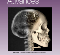
September 28, 2015 — Adult survivors of childhood brain tumors have lower working memory performance compared to healthy adults, according to researchers at Georgia State University and Emory University using functional magnetic resonance imaging (fMRI).
The findings, published in the Journal of the International Neuropsychological Society in August, report that adult survivors of pediatric posterior fossa brain tumors performed significantly lower than controls on standardized clinical tests of working memory performance administered in the study.
The researchers studied the working memory of adult survivors of childhood posterior fossa brain tumors versus a healthy control sample using fMRI and neuropsychological measures. Each group consisted of 17 participants.
During fMRI, the participants completed a measure called the n-back task. They were asked to monitor a series of letters and respond “yes” or “no” with their index or middle finger on a button box if an item was presented “n” items before, ranging from one to three letters back. Accurately recalling a letter two or three letters back represented higher working memory capabilities. Participants also completed other standardized clinical measures.
Whole-brain fMRI analyses also found survivors had significantly greater blood-oxygen level dependent (BOLD) activation in the left superior/middle frontal gyri and left parietal lobe of their brain during a verbal working memory task, demonstrating higher activation in these structures. Analyses revealed higher levels of activations in prefrontal regions were associated with lower behavioral performance on higher-load working memory tasks.
“Our goal was to identify the neural mechanisms underlying working memory difficulty in adult survivors of childhood brain tumors,” said Tricia King, Ph.D., associate professor of psychology and neuroscience at Georgia State. “The results suggest that adult survivors of pediatric posterior fossa brain tumors recruited additional resources to control cognitive ability in the prefrontal lobe during increased demands for working memory. This increased prefrontal activation is associated with lower working memory performance.”
Adult survivors of childhood brain tumors are at risk for neurocognitive deficits, such as working memory impairment, that contribute to poor long-term outcomes. While advances in diagnosis and treatment have led to improved clinical outcomes and increases in the five-year survival rates of pediatric brain tumor patients, research has shown that long-term childhood brain tumor survivors suffer from adverse health, disrupted quality of life, and impaired cognitive and social outcomes.
Working memory deficits are also common in other neurological conditions, such as schizophrenia, multiple sclerosis and traumatic brain injury, because working memory is an essential component for higher-order cognitive processes in humans.
Understanding of the neural mechanism underlying working memory impairments in adult survivors of childhood brain tumors is limited and little fMRI research with these survivors has been reported. This study was designed to address this gap in knowledge and improve treatment for survivors of childhood brain tumors.
Collaborators for the project include Sabrina Na of Georgia State and Hui Mao of Emory University. The study was funded by the American Cancer Society’s Research Scholar Grant (King) and a Brains and Behavior graduate student fellowship (Na) from Georgia State.
For more information: www.psychology.gsu.edu

 April 04, 2024