January 2, 2019 — Imagine an atlas containing an image bank of the blood vessels of the brain taken from healthy humans which can be used as a reference to target alterations among those with cerebral vascular degeneration. That key to improving the diagnosis of neurodegenerative diseases and to understanding their origin is now within reach as the result of the work of a team of Université de Sherbrooke researchers that is gaining international attention.
Kevin Whittingstall, a research professor at the faculty of medicine and health sciences (FMSS) of the Université de Sherbrooke and at the Centre de recherche du CHUS, has dedicated years to develop noninvasive imaging techniques to view the structure and functioning of the human brain. His doctoral student, Michaël Bernier, who is now a postdoctoral fellow at Harvard Medical School, developed a new segmentation software tool. Together, they were able to extract images of blood vessels in the brain that are normally difficult to view using noninvasive procedures. Thanks to these images, they have documented what they call the world's most complete arterial and venous atlas of the human brain. Stephen Cunnane, a professor and researcher at the FMSS and at the Centre de recherche sur le vieillissement, also collaborated in this discovery.
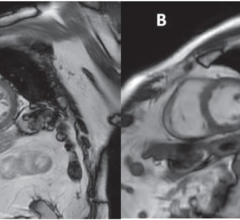
"We spend 10 minutes in a magnetic resonance imaging (MRI) session to take two images of the patient's brain," explained Whittingstall. "The first image targets the arteries by measuring blood velocity. The second image measures magnetic interference, thus enabling the veins to be viewed. Our images are so precise that they enable us to quantify the structure, length and diameter of different blood vessels in each region of the brain. Michaël then assembles them using computer software which combines mathematics with image processing before obtaining an arteriovenous tree that is unique to each patient."
The brain atlas can thus be compared to a fingerprint databank. The team can compare the patient's vascular tree with images taken from healthy brains and determine whether there are subtle variations. If so, the team will try to determine where they originate. Are they from a brain concussion or from the onset of Alzheimer's disease, and so on?
Last November, this innovation made the front page of Human Brain Mapping.1 The team is already being solicited by researchers in other countries who wish to use the atlas in clinical studies on brain concussions and stroke (cerebrovascular accidents).
For more information: www.usherbrooke.ca
Reference
1. Bernier M., Cunnane S.C., Whittingstall K. The morphology of the human cerebrovascular system. Human Brain Mapping, Sept. 28, 2018. https://doi.org/10.1002/hbm.24337

 April 24, 2024