As soon as the Compressed SENSE technology became available to the MRI team at Kantonsspital Winterthur (Switzerland), the site started applying the acceleration possibilities for their MRI scans of the brain, spine and joints, as well as pelvis and abdomen. Using Compressed SENSE appeared a simple yet powerful way to accelerate MRI scanning for different contrast types and sequences, in 2-D as well as 3-D. The significantly reduced scan times convinced the team to plan for adjusting all their ExamCards, with the objective to shorten patient timeslots and then actually plan for increasing their number of MRI patients per day. This is of strategic importance to help them generate revenue when reimbursements go down.
How Fast Can We Scan Without Losing Image Quality?
The new Philips Compressed SENSE technology is a powerful acceleration technique for a wide variety of MRI sequences in a broad range of anatomies. The method combines compressed sensing and sensitivity encoding as in SENSE into one, more powerful, acceleration technique called Compressed SENSE.
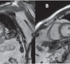
Kantonsspital Winterthur (KSW) was one of the first 10 sites in the world to receive Compressed SENSE on its Ingenia 1.5T and Achieva 3.0T MRI scanners. Sartoretti and radiologist Rene Patzwahl, M.D., have been using it since September 2017 and are very satisfied with the results. “As Compressed SENSE works in virtually all anatomical areas and with many different scan techniques and contrast types, it has the potential to help us reduce the scan time per patient. We can, for example, accelerate a routine brain protocol with six sequences (DWI, transverse T2, transverse T1 IR, SWIp, 3D FLAIR, 3D T1 TFE with gadolinium) by 22 percent by accelerating three sequences (SWIp, 3D FLAIR, 3D T1 TFE with gadolinium) between 30 and 40 percent. That shorter scanning time will then benefit our patients and in addition, it will allow us to scan more patients,” said Sartoretti.
In order to get there, Compressed SENSE has to be incorporated into KSW’s customized set of ExamCards. “Our goal is to reduce scan time, but we want the same image quality as before, because most of our ExamCards have been meticulously optimized to our preferences,” she continued.
“Our approach for the last weeks has been to add an additional sequence with Compressed SENSE to the original exam, and then compare the images,” concluded Sartoretti. In this approach we have initially been using a Compressed SENSE (CS) factor as recommended by Philips, then followed by applying either higher or a lower CS factors. Repeating this in the next patient and for the different sequences, and carefully looking at the images afterwards, helps us decide what the best Compressed SENSE factor is for us.”
*Compared to scans without compressed SENSE. Results from case studies are not predictive of results in other cases. Results in other cases may vary.
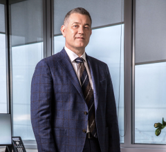
Sabine Sartoretti, M.D., is head of neuroradiology at the Institute of Radiology and Nuclear Medicine, Kantonsspital Winterthur in Winterthur, Switzerland. She specializes in diagnostic neuroradiology. Her interests are dedicated stroke imaging, intra- and extra-cranial vessel wall imaging, brain tumor imaging, perfusion weighted imaging, diffusion weighted imaging and MR spectroscopy.


 April 17, 2024