October 10, 2017 — Elekta announced that members of the Elekta MR-linac Consortium reported data related to the advancement of the company's magnetic resonance/radiation therapy (MR/RT) system at the American Society for Radiation Oncology (ASTRO) Annual Meeting, Sept. 24-27 in San Diego.
The Elekta MR-linac is the only MR/RT system, according to the company, that integrates a high-field (1.5 Tesla) MRI scanner with an advanced linear accelerator and intelligently-designed software. The system is expected to deliver precisely targeted radiation doses while simultaneously capturing the highest-quality MR images. These features are expected to allow clinicians to visualize tumors at any time and adapt treatment.
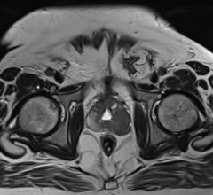
Researchers at the Odette Cancer Centre, Sunnybrook Health Sciences Centre in Toronto, presented preliminary results of a study evaluating the potential of MR-linac in adapting to changes in tumor volume that can be observed using MRI during treatment of glioblastoma multiforme, an aggressive form of brain cancer (Abstract #3706). The study is designed to capture MR images from 20 patients at the treatment planning stage, at 10 and 20 days of radiation therapy, and at one month following completion of treatment. Imaging data showing relative and absolute changes in gross tumor volume (GTV) are then used to simulate a new planning target volume (PTV) to assess the feasibility of treatment plan adaptation.
Data for the first three patients evaluated in this study were presented by Sunnybrook's Mark Ruschin, Ph.D., MCCPM. Two of the three patients showed decreases in GTV of 14 percent and 20 percent at Day 20 and 51 percent and 42 percent one month following completion of treatment. The image set containing the 20 percent GTV shrinkage at Day 20 was used to generate an adapted plan, resulting in a new PTV that was 31 percent smaller than the original. In the adapted plan, the smaller PTV contributed to a reduction in the maximum dose to the brainstem and optic chiasm by 37 percent and 39 percent, respectively. In addition to demonstrating a dosimetric advantage of adaptive brain radiotherapy, the present work is an important first step towards developing the tools and processes needed for clinical implementation of the new MR/RT technology. Future work involves the use of functional imaging to improve the quantification of tumor and normal tissue response to radiation and ultimately leads toward adapting treatment based on an individual patient's biological response.
"Reducing treatment volumes and sparing healthy tissue is a critical factor in improving the care and outcomes for patients treated with radiation therapy," said Arjun Sahgal, M.D., deputy chief of the Department of Radiation Oncology at Sunnybrook. "These preliminary data suggest that the integrated, high-field MR imaging capabilities of MR-linac could enable online plan adaptation in response to changes in tumor volume and surrounding structures during treatment. These adaptations could enable reduced treatment volumes and protection of critical organs and structures."
The potential of MR/RT to provide greater insights into the biology of cancer was also featured at the conference. Allen Li, Ph.D., professor and chief of medical physics at Froedtert & Medical College of Wisconsin moderated and participated in a panel discussion titled "MRI-guided adaptation: From anatomy to biology" (Panel 04). The discussion focused on recent advances in using MRI to assess treatment response and guide adaptive radiation therapy practices, including: a review of data that provide insight into using advanced MRI technologies to assess or monitor radiation therapy responses in selected tumor sites; algorithms and tools required for treatment plan adaptation based on treatment responses; and opportunities and challenges related to the use of advanced MRI in clinical practice.
Additional abstracts related to the development of MR-linac presented at the conference include:
- Quantifying complex abdominal organ motions in different time frames in radiation therapy (Abstract #3669)
This abstract reports results of a study that investigated abdominal motion induced by respiration and peristalsis during various time durations relevant to radiation therapy. The study used a variety of computed tomography (CT) and MRI techniques to acquire images of tumors and surrounding tissue in 31 patients with pancreatic or liver cancer. Images were captured while patients engaged in free breathing (FB) and breath hold (BH). Investigators conclude that the abdominal motions due to peristalsis in the time frames from seconds to minutes can be similar in magnitude to motion resulting from breathing. These motions can be irregular and persistent throughout the imaging and radiation therapy delivery procedures, and should be considered together with respiration motion during radiation therapy for abdominal tumors.
- Acceleration of online adaptive replanning with workflow automation (Presentation #238)
This abstract reports on the development of software tools to automate online adaptive re-planning (OARP) and the testing of these tools on both a conventional linear accelerator with an in-room CT scanner and an MR-linac. The research treatment-planning package has the ability to account for magnetic fields and includes fast replanning algorithms. The OARP process was broken down into five distinct modules: auto-segmentation based on daily CT or MRI scans; contour review and editing; adaptive plan generation; evaluation of the adaptive plan based on daily images; and transferring the new plan and performing quality assurance assessment prior to delivery. The automated tools and workflow were tested on 23 daily CT and MRI scans from five prostate cancer cases. Results show that OARP with the automation software tools, excluding the contour editing module, was 250 ±14 seconds, a 49 percent reduction in operation time compared with OARP conducted without the automated tools. The contour editing module, which was the most time consuming, ranged from five to ten minutes. Investigators conclude that the use of these automation tools substantially reduces the time for the online replanning process and eliminates user stresses and input errors during the online replanning process. The research team is working to improve the efficiency of the contour editing module.
The founding members of the Elekta MR-linac Consortium are:
- University Medical Center Utrecht, the Netherlands (UMCU);
- The Netherlands Cancer Institute-Antoni van Leeuwenhoek Hospital, Amsterdam, the Netherlands;
- The University of Texas MD Anderson Cancer Center, Houston, Texas;
- The Institute of Cancer Research, working with its clinical partner The Royal Marsden NHS Foundation Trust, London, England;
- Froedtert & the Medical College of Wisconsin Clinical Cancer Center at Froedtert Hospital, Milwaukee, Wisconsin;
- The Christie NHS Foundation Trust, Manchester, U.K.; and
- The Odette Cancer Centre, Sunnybrook Health Sciences Centre in Toronto.
Elekta's MR-linac is a work in progress and not available for sale or distribution.
For more information: www.elekta.com

 April 24, 2024