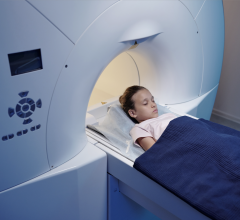
One of the biggest hurdles of hybrid positron emission tomography and magnetic resonance (PET/MR) imaging is the prevalence of motion artifacts—blurring and ghostly visual anomalies caused by patient motion on the table during imaging. An MR technology has now been designed for PET/MR that employs tiny radiofrequency solenoids—metal coils integrated into hardware placed on the body—to track motion from those who do not or cannot stay put. Special software can then use the additional information provided by the coils to optimize the image, according to research being revealed at SNMMI’s 2013 Annual Meeting.
One of the most important applications of this technology is in brain imaging for patients with dementia, who often have movement disorders such as those in Parkinson’s. For these patient populations, this technology improves already exceptional diagnostic imaging and creates better potential for therapy monitoring in the future.
“Dementia is one of the biggest health problems facing human society now and in the future. PET is a powerful tool in early detection and treatment of dementia, including Alzheimer’s disease. Early diagnosis of dementia can have a tremendous impact on treatment for patients and their family members,” said Chuan Huang, Ph.D., lead researcher for the study, from Massachusetts General Hospital in Boston, Mass. “Simultaneous PET/MR allows measurement of anatomy, functionality, and biochemistry of tissues and cells. Combined with our low-cost MR micro-coil–based motion correction, PET/MR provides essential information about the brain and other parts of the body with greater accuracy, even for long studies or research involving motion, which opens the door to more expansive multi-modality studies.”
During brain scans in particular, patient head motion without corrective technology can lead to imaging failures due to extensive blurring in reconstructed PET data. Even patients who are restrained are likely to move position during scans that can take an hour or more, and just a few millimeters or degrees off can cause artifacts.
“PET imaging is similar to taking a photo inside the body using specialized imaging data,” said Huang. “Just like with a normal camera, when you take a picture in an extremely lowlight environment you need long exposure times to get a good quality picture. If the object is moving, the image blurs. Our research is essentially providing an image stabilizer for PET/MR scanners, which can then generate crisp and clear images even though the object is moving. Our approach is similar to capturing each of the light rays coming from the moving object and configuring it back to its original position.”
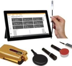
The micro-coils are smaller than a dime and just millimeters in diameter. Before scanning they are fixed on an apparatus placed on the patient’s body, providing real-time and 3D fields of motion during imaging that are incorporated during image reconstruction. Other motion tracking techniques already in clinical use include “gating” technologies that involve the equivalent of freezing a motion picture and arranging film stills in a pattern that omits motion, then splicing them back together; but this means effectively throwing away meaningful information between data splices, which degrades scan quality.
Researchers conducted the study by using phantoms, or mock humans, and motion was simulated using a ventilator system. The MR micro-coil apparatus was positioned on the phantom, and motion tracking was acquired simultaneously during PET/MR scanning. Results of the study showed that 3D motion-tracked PET imaging using MR micro-coils dramatically reduced movement-related imaging artifacts. Now that this PET/MR technology has been proven beneficial, further research will be needed with actual patients before it can be expanded into general imaging practice.
In addition, the researchers are developing wireless micro-coils for use with PET/MR. “The wireless micro-coils are, compared to the wired coils, more patient friendly, easier to set up and cheaper to manufacture,” added Jinsong Ouyang, Ph.D., senior researcher for the study, also from Massachusetts General Hospital in Boston, Mass. “The advantages should smooth adoption of motion-tracking micro-coils in clinical practice.”
Scientific Paper 44: Chuan Huang, Yoann Petibon, Thomas Brady, Georges El Fakhri, Jinsong Ouyang, Center for Advanced Medical Imaging Sciences, NMMI, Radiology, Massachusetts General Hospital, Harvard Medical School, Boston, MA; Jerome Ackerman, Martinos Center for Biomedical Imaging, Massachusetts General Hospital, Harvard Medical School, Boston, MA, “Real-time 3D motion tracking using MR micro-coils for PET imaging,” SNMMI’s 60th Annual Meeting, June 8–12, 2013, Vancouver, British Columbia.
For more information: www.snmmi.org

 April 10, 2024


![(A) PET images of [68Ga]Ga-DOTA-ZCAM241 uptake at baseline and 3, 7, and 12 days after injection as inflammatory arthritis developed in single representative individual mouse. Images are normalized to SUV of 0.5 for direct comparison between time points. (B) CD69 immunofluorescence Sytox (Thermo Fisher Scientific) staining of joints of representative animals during matching time points.](/sites/default/files/styles/feed_medium/public/PET%20Tracers.jpeg?itok=P5Di6MIe)