January 28, 2015 — Using a camera and water tank, investigators from Dartmouth-Hitchcock’s Norris Cotton Cancer Center, led by Brian Pogue, Ph.D., and David J. Gladstone, ScD, demonstrated that induced Cherenkov light can be imaged and used to confirm that the complex spatial dose distribution imparted in dynamic treatment plans is being delivered correctly. The paper “Video-rate optical dosimetry and dynamic visualization of IMRT and VMAT treatment plans in water using Cherenkov radiation,” was published in Medical Physics.
“Cherenkov radiation is emitted and exists as a ‘free’ optical signal that has not been previously utilized,” explained Adam Glaser, lead Ph.D. student on the project. “By imaging the light, we can improve the quality and accuracy of delivered plans to prevent radiation mistakes. This will ultimately improve the overall outcome for patients being treated.”
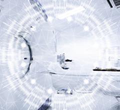
The Cherenkov Effect is responsible for the characteristic blue glow of nuclear reactors. Although the phenomenon has been constructively utilized for decades in high-energy particle physics and astrophysics, only recently has there been investigation into its utility in radiation therapy. In that context, it is the only current method that can reveal dosimetric information while remaining completely non-invasive. Essentially, the signal comes directly from the water phantom being irradiated rather than introducing detector arrays.
To accomplish this work, a high-sensitivity, intensified CCD camera was set up to take a two-dimensional projected image during delivery of the Intensity-Modulated Radiation Therapy (IMRT) or Volumetric Modulated Arc Therapy (VMAT) plans. The camera focused on a water bath, and was gated to the Linear Accelerator to take a rapid series of photos that were connected to make an ersatz video and used to compare overall light distribution with the original dosing plan.
“Compared to conventional [quality assurance] methods, the advantages of this technique are video-rate, real-time data acquisition, two dimensional projection of 3-D dose distribution, and high spatial resolution that is limited only by the collection optics of the system,” said David J. Gladstone, ScD.
Next steps for the team at Norris Cotton Cancer Center are to extend the system to tomographic 3-D data recovery, as well as continuing clinical trials to investigate Cherenkov light imaged directly from a patient’s tissue surface.
For more information: www.cancer.dartmouth.edu
 March 28, 2024