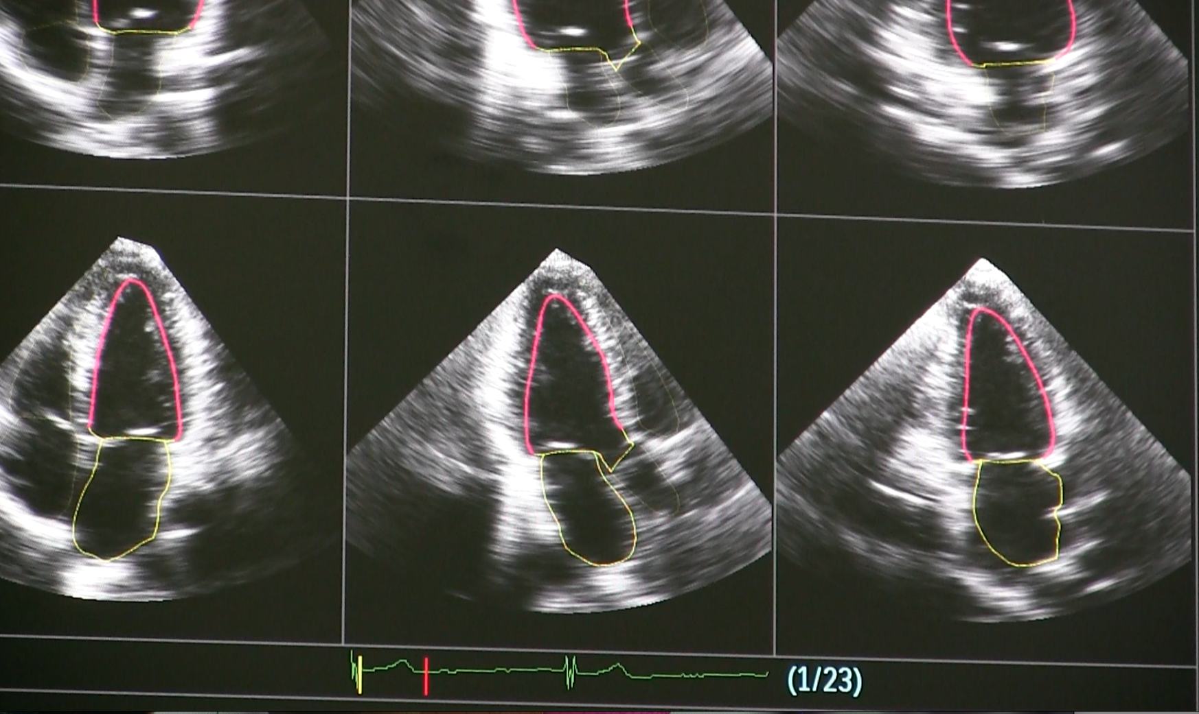
The Philips' Epiq ultrasound platform uses artificial intelligence to automate standard echo views of cartdiac anatomy without human intervention. The idea is to eliminate sonographer variability. Image courtesy of Philips
The following is a roundup of a few of the major advances in ultrasound imaging technology over the last 12 months.
Automated Breast Ultrasound
For the last decade or so, women’s health professionals and advocates have worked to educate the public about the role that dense fibroglandular breast tissue plays in breast cancer risk. Women with dense breasts are at higher risk of having cancers missed on their mammogram because the tissue masks lesions and makes them difficult to detect. As a result, those on the forefront of women’s health are moving beyond the view of mammography as a one-size-fits-all solution for breast cancer screening and urging women to discuss supplemental screening options with their physicians. Breast ultrasound has emerged as an option for women with dense breasts, and several studies have shown that it can detect cancers that were initially missed on mammography.
In September, Delphinus announced enrollment of the first patient in its own clinical trial comparing the efficacy of its SoftVue 3-D whole breast ultrasound system to digital mammography. The company hopes to enroll 10,000 asymptomatic women with dense breast tissue in the SoftVue Discover Breast Ultrasound Prospective Case Collection project, with the goal of assessing the ability of SoftVue to detect additional cancers. Data from the study will be included in Delphinus’ submission of a U.S. Food and Drug Administration (FDA) premarket approval (PMA) application for a supplemental screening indication for women with dense breasts in combination with mammography.
The fully automated system performs a 360-degree scan of the breast while patients are in the prone position. This approach addresses several of the pitfalls of breast exams with conventional handheld ultrasound systems:
• Automating the scan eliminates the chance of critical findings being missed due to operator variability;
• Automation significantly reduces the time needed to complete the exam, cutting the procedure to 2-4 minutes; and
• Conducting the scan in the prone position increases patient comfort for an exam that many women find uncomfortable.
“Dense breast tissue can mask or hide cancer, making it more difficult for mammography to detect cancer. And while ultrasound has been shown to be effective in detecting cancer in dense breasts, there’s a need for advanced technology like SoftVue that enables fast and comfortable whole breast ultrasound with fewer false positives,” said Mary Yamashita, M.D., assistant professor of clinical radiology at the University of Southern California Norris Comprehensive Cancer Center and the national principal investigator of the Discover Breast Ultrasound project.
OB/GYN Ultrasound
Philips Healthcare in September introduced new features to enhance OB/GYN ultrasound exams on its Epiq 7 and 5 and Affiniti 70 ultrasound systems. The guiding principle of the new features is to give clinicians more information earlier in pregnancy and hopefully allow more treatment options to deal with potential challenges.
Philips had previously introduced Anatomical Intelligence technology on its cardiovascular ultrasound systems to automate structural measurements. The new aBiometry AssistA.I. feature plays a similar role, preplacing measurement cursors on selected fetal anatomy structures. This reduces the number of steps associated with conventional measurements and streamlines reporting workflow.
The 3D9-v3 transducer now sports a 2-D Tilt feature that allows for lateral scanning of anatomical structures that are off-axis without having to manually angle the transducer, making the exam more comfortable for the patient.
The new eL18-4 transducer features a multi-row array configuration for full electronic focusing of the elevation plane to provide exceptional detail resolution and tissue uniformity.
Cardiovascular Ultrasound
The rapid advancement of technologies to solve structural heart issues has greatly enhanced the value of ultrasound as a tool for procedural navigation in the OR and the cath lab. Such procedures require highly detailed, real-time imaging to precisely place devices or guide surgical interventions.
Siemens Healthineers received FDA clearance in September for a new feature on its Acuson SC2000 premium ultrasound system that was designed specifically to aid in structural heart interventions. TrueFusion marries real-time soft tissue and blood flow information from the SC2000’s True Volume transesophageal echo (TEE) transducer with 2-D fluoroscopy imaging from Siemens’ Artis with Pure angiography system to provide both anatomical and functional markers for the procedure. This integrated approach helps improve communication between interventionalists and echocardiographers, which reduces procedure times and improves patient outcomes. TrueFusion is available on Release 5.0 of the Acuson SC2000.
Toshiba Medical’s Aplio i900, which received FDA clearance in March, is another example of an ultrasound system that can be used for interventional cardiac procedures. The latest addition to the company’s premium Aplio i-series is powered by iBeam beam-forming technology, which optimizes the efficiency of the ultrasound beam to improve image quality through enhanced penetration and spatial and contrast resolution. Toshiba also focused on making the system ergonomic and user-friendly with iSense technology and touch control screens for streamlined workflow; real-time quick scans let users make automatic image adjustments without pushing a button. The company said this combination of enhancements gives healthcare providers a cost-effective premium solution for everyday clinical settings.
Point-of-Care Ultrasound
Clarius Mobile Health introduced new advanced features over the summer for its C3 and L7 wireless handheld ultrasound scanners, which wirelessly send data to most iOS or Android smart devices. This gives clinicians the capability to perform quick scans virtually anywhere with commercially available devices. The Clarius C3 multipurpose scanner is designed to image the abdomen and lungs, with a virtual phased array for quick scans of the heart. The Clarius L7 specializes in procedure guidance and imaging of superficial structures.
The new features are part of the Clarius App 3.1 Eclipse, which gives users enhanced capabilities for annotations, automated heart-rate monitoring during cardiac scanning and easy bladder volume measurement. The devices can be used in an educational setting as well thanks to a new feature that allows sharing of imaging screens with multiple devices simply by scanning a QR code.
Read the related article "Recent Advances in Echocardiography Technology."

 April 17, 2024