February 5, 2013 — A new computed tomography (CT) scanner substantially reduces potentially harmful radiation while still improving overall image quality. National Institutes of Health (NIH) researchers, along with engineers at Toshiba Medical Systems, worked on the scanner. An analysis of data on 107 patients undergoing heart scans found that radiation exposure was reduced by as much as 95 percent compared to the range of current machines, while the resulting images showed less blurriness, reduced graininess and greater visibility of fine details.
The machine recently received approval by the U.S. Food and Drug Administration (FDA), but more studies will be needed before it can be adopted for wide clinical use.
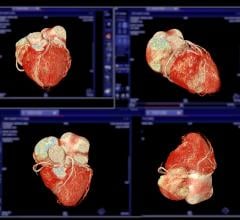
“CT scans are a great diagnostic tool for heart disease because we can obtain high-resolution 3-D images of the heart quickly and non-invasively,” said coauthor Andrew Arai, M.D., chief of the Cardiovascular and Pulmonary Branch at the NIH’s National Heart, Lung and Blood Institute (NHLBI). “However, the benefits of CT have been tempered by concerns over the radiation required to achieve these images. With this next-generation device, we are close to achieving the best of both worlds.”
Most CT scanners available in clinics have 64 rows of X-ray detectors. The new scanner has 320 detector rows, which allow imaging of a larger area of the body at one time. The new scanner also has a more powerful X-ray beam generator. And the gantry — the doughnut-shaped part of the CT machine — can complete a full rotation in 275 milliseconds. Current scanners top out at 350-millisecond rotations. In addition to hardware advances, the NHLBI team worked on the device settings and features with Toshiba to optimize radiation usage and image quality.
“These multiple advancements work together to allow us to image the entire heart within one heartbeat about 93 percent of the time,” noted lead study author Marcus Chen, M.D., a clinician in the NHLBI’s Advanced Cardiovascular Imaging Laboratory.
“These improvements could help clinicians identify problems in even the smallest blood vessels or enable them to conduct complicated tests like measuring blood flow in the heart while limiting radiation exposure,” Chen added.
Between July and October 2012, Chen and colleagues used the new scanner to perform CT angiographies — which look for plaque buildup or other problems in the coronary arteries — on 107 adults of varied height and weight, between the ages of 27 and 82. The research team then compared both the radiation dose and image quality of the new CT scans to 100 scans taken on a first-generation 320-detector row scanner at the NIH campus between January and April 2010.
The median effective radiation dose for the new scanner was 0.93 millisieverts (mSv), compared to 2.67 mSv for the first-generation scanner, and almost every patient (103 of 107) received less than 4 mSv of radiation. (millisieverts reflect how much radiation a body absorbs, so it can help determine potential health risks. The average person receives about 2.4 mSv of background radiation each year.) Nationwide, coronary CT angiography typically involves effective radiation doses between 5 and 20 mSv, depending on the patient's body type and the quality of the machine.
The study, which was published Jan. 22 online in the journal Radiology, was funded by the NHLBI intramural research program (HL006138-02). The CT machine was provided to the NHLBI by Toshiba Medical Systems through a cooperative research agreement.
The National Heart, Lung, and Blood Institute (NHLBI) is a component of the National Institutes of Health. NHLBI plans, conducts, and supports research related to the causes, prevention, diagnosis and treatment of heart, blood vessel, lung and blood diseases; and sleep disorders. The Institute also administers national health education campaigns on women and heart disease, healthy weight for children, and other topics.
For more information: www.nhlbi.nih.gov

 April 17, 2024