June 20, 2013 — A relatively new weapon in the fight against childhood brain cancer has emerged that improves upon standard magnetic resonance imaging (MRI) by providing information about tumor metabolism and extent of cancer in children diagnosed with glioma, a growth caused by the abnormal division of glial cells in the brain, said researchers at the Society of Nuclear Medicine and Molecular Imaging’s 2013 Annual Meeting.
Brain cancer imaging is often conducted with conventional MRI, but there are some limitations to this imaging technique. This type of cancer accounts for approximately 80 percent of all invasive brain tumors and develops in the brain’s glial cells that protect and maintain a state of neural equilibrium. Conventional MRI can sometimes over- or underestimate the extent of these tumors and the exact shape of their outlying margins. A method of molecular imaging called positron emission tomography (PET), which provides information about physiological functions rather than structures of the brain, can be performed with a variety of imaging agents that bind to specific cellular systems to image gliomas. Two of the main types of brain imaging agents used for this purpose provide information about either glucose, or sugar, metabolism of cells or the cellular metabolism of amino acids, the brick-and-mortar components of proteins used by tissues, especially rapidly growing so-called neoplastic tumors.
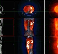
Amino acid PET imaging has been shown to be better for detection of neoplastic tissue and treatment monitoring in cases of brain cancer than glucose imaging. In general, the brain requires more glucose than the body’s other tissues and organs, making brain scans “noisier” and less defined than others due to this increase in overall cellular metabolism, whereas areas of increased amino acid activity show up clearly on scans as a visual “hot spot.” This study focuses on a particular amino acid imaging agent, O-(2-[18F]-fluoroethyl)-L-tyrosine (F-18 FET), and its diagnostic benefit for imaging pediatric gliomas when conventional MRI cannot make out a clear picture of disease.
“Cancer of the brain and spinal cord are common in children and young adults, and caring for this particular group can be challenging because choice of treatment depends on specific information about the tumor. Tumors in younger patients show a greater variety in both type and size, and in many cases the tumors are located near critical brain structures that prohibit surgical removal,” said Veronika Dunkl, M.D., a research scientist at the Institute of Neuroscience and Medicine, Forschungszentrum Jülich, in Jülich, Germany. “In patients with brain tumors, contrast-enhanced structural MRI is currently the diagnostic method of choice. However, in youths with newly diagnosed cerebral lesions thought to be brain tumors, MRI’s ability to identify neoplastic tissue or tumor progression and recurrence after treatment is limited. F-18 FET is complementary and can potentially improve diagnosis and treatment of pediatric brain tumors.”
Pediatric brain imaging with PET and F-18 FET can be used not only to evaluate extent of tumors but also to help doctors plan for biopsy, surgery and radiation therapies and track response to therapy and recurrence of tumors after completion of a treatment cycle.
F-18 FET is also unique from other amino acid PET agents because the production of the drug can be centrally located and distributed by a radiopharmacy, whereas other amino acid–based PET agents must be produced by an on-site cyclotron — a massive particle accelerator that bombards particles with a target used to radiolabel the agent’s molecular compound. For F-18 FET, it is the amino acid tyrosine that allows brain cells to signal each other. The greater logistical ease of F-18 FET is due to its radioactive half-life of approximately 110 minutes, whereas many other isotopes have a half-life of only about 20 minutes and must be administered for patient imaging almost immediately.
For this study, 15 young patients suspected of glioma cerebral cancer via MRI screening underwent PET imaging with the guidance of F-18 FET. This molecular imaging technique was found to be highly effective, about 87 percent, for detecting and differentiating brain lesions in children and young adults. The method was able to pinpoint 11 out of 12 brain lesions correctly as tumors and two out of three as a non-tumorous growth. Repeated PET imaging (17 scans) for seven more pediatric patients provided meaningful information about cancer progression or remission. F-18 FET imaging was able to detect residual tumor and tumor progression in five out of six scans, and in 11 scans in which the cancer had been eradicated, for a 94 percent rate of accuracy.
“Results of the present study may improve the clinical management of this vulnerable patient population significantly, especially when a decision for further treatment is difficult or impossible on the basis of conventional MRI alone,” said Dunkl.
The National Cancer Institute estimates that brain cancers are among the most common cancers in child populations. Incidence of pediatric brain tumors is approximately 3.2 cases per 100,000 people.
Scientific Paper 474: Veronika Dunkl, Gabriele Stoffels, Gereon Fink, Nadim J. Shah, Heinz Coenen, Karl-Josef Langen and Norbert Galldiks, Institute of Neuroscience and Medicine, Forschungszentrum Jülich, Jülich, Germany; Corvin Cleff, Department of Neurology, University of Cologne, Cologne, Germany; and Sevgi Sarikaya-Seiwert, Department of Neurosurgery, University of Düsseldorf, Düsseldorf, Germany, “The use of dynamic O-(2-[18F]fluoroethyl)-L-tyrosine-PET in the clinical evaluation of brain tumors in children and young adults,” SNMMI’s 60th Annual Meeting, June 8–12, 2013, Vancouver, British Columbia.
For more information: www.snmmi.org

 April 05, 2024