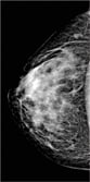
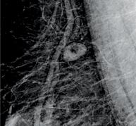
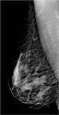
Right craniocaudal (RCC) Photos courtesy of Philips Healthcare
United Medical Center (UMC) is a non-profit, full-service community hospital serving Southeast Washington, D.C. and nearby Maryland communities. Though rich in African-American heritage, the community faces many economic challenges.
UMC, an independent government instrumentality in eyesight of the U.S. Capitol, cares for patients with a multitude of health problems and little access to quality healthcare. The hospital aims to provide its community with the best care possible, offering full-service solutions from pediatrics to women’s health, recently renewing its women’s health offerings. For years, UMC’s radiology department used an analog mammography system to administer mammograms to its patients. In an effort to modernize the radiology department and provide better, more efficient care for its patients, UMC took the leap of replacing its analog mammography system with a digital solution.
Choosing the Digital Mammography Solution
Until recently there was only one digital mammography choice. UMC’s decision to transition to a digital mammography system was timely due to the addition of a new digital mammography solution in the United States. The Philips MicroDose mammography system, which has been available in Europe and Canada for numerous years, recently made its entry to the U.S. market. The UMC team immediately was intrigued because it offered a lower radiation dose than other standard full-field digital mammography systems, while maintaining high diagnostic quality images.
The MicroDose digital mammography system’s technology counts individual X-ray photons using a 50 micrometer detector element, creating very low noise and eliminating analog to digital conversion. By using matching pre- and post-collimators, only those X-rays perfectly aligned with the detector are allowed to pass through the breast, eliminating 97 percent of scatter and yielding high quality images. (See Figure 1.)
A study of breast radiation exposure recently published by the Irish Breast Screening Program compared radiation dose to the breast per exposure and per exam among the digital mammography systems of three leading vendors. The study concluded that Philips Healthcare’s MicroDose low-dose mammography system had the lowest mean glandular radiation dose per image and per exam among the three vendors. (See table below.)
Reduced radiation dose was an information factor in UMC’s decision-making process. The tomosynthesis mammography solution uses more radiation dose, which the center tries to minimize for its patients. In choosing its system, UMC wanted to provide a safer solution for patients, especially those who avoid getting exams due to fear of radiation exposure. The solution also improves clinical throughput by providing faster exams — five minutes from scan to image acquisition, which was a key factor, as the center administers up to 200 mammograms per month.
Converting From Analog to Digital
Since UMC is a safety net hospital, it initially was a struggle to obtain the hospital’s approval to acquire the MicroDose system, due to its lean investment in new equipment. Once the health benefits for patients was explained to hospital administrators, in addition to improved throughput with a single technologist and removal of the film processor, toxic chemicals and plumbing concerns, the approval process became a lot easier. The new digital mammography system also provided the final component to have a 100 percent American College of Radiology (ACR) accredited facility, the first time in the history of the hospital.
After obtaining the approval for the system, the transition from analog to digital was seamless and easy. Prior to installing the digital mammography solution, UMC’s mammography suite was an ad hoc patchwork of rooms inherited from years of renovation and configuration changes. The mammography suite left no space unused. The waiting room, processor, dark room and analog system filled a tight space. Once the processor room, plumbing and storage bins for films were removed, the space was reconfigured with a cooling room for the electronics. From the technologists’ perspective, the conversion to a digital mammography solution provided enhanced throughput, eliminated the need for film manipulation, cassettes, toxic photographic liquids and mechanical processors. From the radiologists’ perspective, the streamlined integration of a digital solution is intuitive with existing plain film, CT and MRI viewing systems, with obvious and familiar benefits as any digital modality.
Clinical Experience With the New Digital Solution
Since the system’s installation in August 2012, telephone calls for information on cancer screening have increased. Patients from other states have sought UMC’s services. Positive feedback was received from patients, who have commented that their exam was efficient and more comfortable, since the warmed, curved surface of the exam table is less intrusive than other standard mammography systems. Also, patients who have had radiation therapy for breast cancer treatment are now less reluctant to get their mammograms due to the lower radiation in MicroDose exams.
As far as the clinicians’ experience, the footprint of the Philips MicroDose system is similar to other vendors — the only difference was a cooled computer closet. Clinical space was not gained in the mammography suite. However, radiologists’ satisfaction with the image interpretation of the digital mammograms is commensurate with the recent 2012 study by Cole, et al.3, which concluded that photon-counting full-field digital mammography was not inferior to conventional digital mammography. The breast imagers at UMC are very pleased with the diagnostic quality images.
Many Benefits
MicroDose completed the total replacement of legacy equipment. The facility passed ACR and U.S. Food and Drug Administration (FDA) accreditation easily. Transitioning from analog to digital mammography has provided many benefits — not only to the hospital, but also to its patients. The installation of the digital mammography system provides a market share advantage in a consumer-driven medical examination. Patients find the exams more efficient and comfortable, while clinicians are impressed with the high quality of the images even at reduced radiation dose. Overall, the solution’s lower radiation dose with the highest resolution provides cache and uniqueness to UMC’s portfolio of imaging services. itn
Raymond Tu, M.D., is chairman of radiology at United Medical Center in Washington, D.C., clinical associate professor of radiology at The George Washington University in Washington, D.C. and partner, Progressive Radiology, at Falls Church, Va. He is chapter president of the District of Columbia chapter of the American College of Radiology (ACR), and served as physician member of the D.C. Board of Medicine and chairs of the ACR Medicaid network. Tu is a member of the Medicaid J12 Carrier Advisory Committee.
Steve Rothenberg, MS-III, The George Washington University School of Medicine, Washington, D.C.
Riad Charafeddine, M.D., is staff radiologist at United Medical Center in Washington, D.C., and radiologist at Progressive Radiology, Falls Church, Va.
Theodore Williams, MA, RT, is director of radiology and cardiology services at the Department of Radiology, Not-for-profit Hospital Corporation dba United Medical Center, in Washington, D.C.
Rory Grace, MS-III, The George Washington University School of Medicine, Washington, D.C.
References
1 Åslund M, Cederström B, Lundqvist M, Danielsson M. “Scatter rejection in multi-slit digital Mammography.” Medical Physics 2006; 33(4):933-40.
2 Baldelli P, McCullagh J, Phelan, N, Flanagan F. “Comprehensive dose survey of breast screening in Ireland.” Radiation Protection Dosimetry 2012; 145:52-60.
3 Cole EG, Toledano AY, Lundqvist M, Pisano ED. “Comparison of radiologist performance with photon-counting full-field digital mammography to conventional full-field digital mammography.” Acad Radiol 2012; 19:916-922.
4 Ojuko JM, Young KC, Burch A. “A survey of patient doses from digital mammography systems in the U.S. from 2007 to 2009.” J. Marti et al. (Eds.): IEWM 2010, LNCS 6136, 365-370, 2010.
5 Philips MicroDose mammography system, technical data sheet, U.S./Canada. 2012.
6 Sectra MicroDose mammography. “Highest image quality, Half the radiation.” 2010.
A Range of Digital Mammography Systems Available
The digital mammography market continues to grow, with several players offering systems. Here are a few of the products currently on the market:
• Aspire HD full-field digital mammography (FFDM) system by Fujifilm Medical Systems U.S.A. is U.S. Food and Drug Administration (FDA)-501(k) cleared, and provides image clarity that enables extraordinary detail of potential abnormalities that help assist in more accurate and reliable diagnoses, plus results in enhanced clinician productivity. The system has a proprietary detector that uses dual-layer amorphous selenium (aSe) and the first use of Fujifilm’s direct optical switching (DOS) technology. This new technology, combined with 50-micron resolution, provides detailed visualization.
• GE Healthcare’s Senographe Essential system uses an integrated image chain. From X-ray tube to detector to image-reconstruction software, every component contributes to high image quality at optimum dose for each patient’s breast composition. The system offers Enhanced Detective Quantum Efficiency (DQE); molybdenum/rhodium dual track tube; Automatic Optimization of Parameters (AOP) that transparently select all exposure parameters based on breast radiological properties; three AOP modes that enable flexibility in dose management; and enhanced conspicuity with Fine View and improved contrast with Premium View.
• The Selenia Dimensions from Hologic delivers sharp images for visualization of the finest details; tomosynthesis (3-D) technology for diagnostic performance with optimal workflow efficiencies; one-touch control for seamless, instantaneous transition between imaging modes, including full-field digital mammography (2-D imaging), tomosynthesis (3-D imaging), or “combo-mode” imaging (2-D+3-D imaging; advanced user tools to simplify operation and enable higher patient throughput; and sophisticated, ergonomic features specifically developed to assure the well-being of the patient.
• Planmed’s Nuance Excel is a full-field digital mammography (FFDM) system, and includes the company’s proprietary MaxView Breast Positioning System for enhanced tissue visibility, and Side Access patient positioning for optimal working ergonomics. It also features a large 24 x 31 cm amorphous selenium (aSe) detector. It is intended for both screening and diagnostic mammography.
• The Mammomat Novation S from Siemens Medical Solutions utilizes the latest full-field amorphous Selenium (aSe) detectors, which convert X-ray energy into an electric charge to help detect small and low-contrast objects, yielding excellent image quality. To achieve optimum dose, it utilizes the same technology as the Mammomat NovationDR - OpComp and OpDose.

 April 16, 2024


