There are currently three major trends in cardiovascular computed tomography (CT) technology — lowering radiation dose, development of myocardial perfusion imaging and the use of CT datasets for procedural planning, especially for transcatheter aortic valve replacement (TAVR).
Lowering, Recording the Dose
“Lately the main topic in the industry has been dominated by dose reduction,” said Guillaume Grousset, Siemens product marketing manager for dual-source CT.
He said cardiac CT has traditionally been a very high-dose imaging procedure. To image the heart, a moving object, with previous generations of CT scanners, several cardiac cycles had to be imaged and the best images stitched together to reconstruct each phase. However, with advances in reconstruction software and higher-slice systems, the dose has been brought down significantly in recent years.
Some of the biggest advances in CT have been in software, speeding workflow, adding more quantification and reducing dose. The software with the most far-reaching impact on CT is iterative reconstruction, which reduces noise while attempting to maintain image quality on lower-dose scans. New software is also being added to systems to accurately record radiation dose, which became a requirement by law in California in July. Other states are looking at similar laws.
Toshiba’s Adaptive Iterative Dose Reduction 3D (AIDR 3D) was released in April. It integrates with Toshiba’s Nema XR 25 dose check software to make users aware of the radiation dose being administered. It includes dose alert and dose notification, addressing the two main components of the Medical Imaging and Technology Alliance’s (MITA) CT Dose Check Initiative, as well as tracking and digital imaging and communications in medicine (DICOM), to further enhance dose awareness.
Philips’ iDose iterative reconstruction software has about 500 installs. Its latest version, iDose4, is available on the iCT, Ingenuity CT and Brilliance 64 scanners. The system lets radiologists personalize image quality based on each patient’s specific needs. The advanced algorithms provide up to 57 percent improvement in spatial resolution at low dose; the majority of factory protocols are reconstructed in 60 seconds or less.
Siemens’ latest Sinogram Affirmed Iterative Reconstruction (SAFIRE) software reduces dose by up to 60 percent compared to previous filtered back projection techniques. The extremely fast reconstruction speed of 20 images per second enables reconstruction of a typical high-resolution thorax examination of 30 cm in 15 seconds. It is available for Somatom Definition Flash, Definition AS and DS.
GE Healthcare’s 510(k)-pending Discovery CT750 HD FREEdom (Fast Registered Energies and ECG) Edition will address issues with coronary motion, high heart rates, calcium blooming, plaque composition and accurate myocardial perfusion. The software will go beyond today’s clinical information by providing plaque material composition assessment and accurate perfusion calculations.
Perfusion Imaging is the Next Step
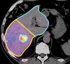
Newer CT software can map the levels of iodine contrast agent present in the heart muscle during each phase of the cardiac cycle. Since the contrast is mixed with the blood, it serves as a surrogate measure of perfusion in the myocardium to help identify coronary artery blockages or infarcts. The current gold-standard for myocardial perfusion imaging (MPI) is single photon emission computed tomography (SPECT), but CT could possibly eliminate the need for the nuclear scan.
“The next logical step is for CT to determine how significant is the stenosis. Is it blocking the flow to the myocardium? What is the severity of the stenosis?” Grousset said.
He said some patients with chest pain stay overnight at the hospital so they can be scanned when the nuclear imaging department opens in the morning. With CT MPI, the results could be seen within minutes of the scan.
CT MPI will take several years to become mainstream, with adoption increasing after a larger amount of clinical validation is acquired. However, CT perfusion imaging is already a well-accepted technique used in neurological imaging for stroke.
CT-Guided Treatment Planning
CT datasets can be sliced and diced to create 3-D images of specific anatomy, which can be used for non-invasive surgical planning and better planning and guidance for interventional procedures. CT imaging has been key to the success of endovascular aortic repair (EVAR) for aortic aneurysms, helping vascular surgeons evaluate access routes, determining the collateral vessels involved and getting precise vessel sizing for proper-fitting stent grafts.
CT also plays a similar critical role for TAVR in assessing access and annulus device sizing. Datasets are also used to precisely determine the location of the aortic root and the right and left coronary arteries, which is critical for device placement. New software uses CT datasets to create overlays on live angiography for better procedural navigation.
CT 3-D renderings are increasingly used as reference images and static overlays on fluoro in cath labs to aid in navigation, especially in tortuous or difficult-to-see anatomy.
Impact of the Economy
CT vendors recognize the slow economy has impacted hospitals. Their focus over the past two years has shifted from showing expensive, latest generation systems to showcasing upgradable and more affordable systems.
A recent example of this trend is Toshiba’s VeloCT console, which allows new enhancements for patient safety, dose management and workflow for existing Aquilion 32, 64 and CX CT systems manufactured between 2006 and 2012. Part of the upgrade allows hospitals to include needed improvements without the vast expense of a new scanner.
The Newest Scanners
In April, Siemens launched the Somatom Definition Flash dual-source system. It uses the new Stellar Detector, the first fully integrated microcircuitry detector that eliminates traditional soldered circuit boards. This greatly reduces electronic noise and increases spatial resolution at lower doses. Grousset said this enables easier visualization of coronary stents with less blooming artifact.
Toshiba’s Aquilion Prime, introduced in January, offers double-slice technology and the coneXact reconstruction algorithm originally designed for Aquilion One. It can generate 160 unique slices per rotation, enhancing multiplanar reformatting (MPR) and 3-D-rendering. It features an 80-row, 0.5 mm detector, a 7.5 MHU large-capacity tube and 0.35-second scanning. This allows shorter scan times. The system also features a 78 cm aperture gantry, the largest available for bariatric patients.
GE released the Optima CT660 in August 2011, a compact system offering ASiR and Snapshot Pulse with adaptive gating to reduce patient dose. It is designed for ease-of-use and is scalable from 32-128 slices through purchasable options. The CT Advantage Workstation cardiovascular system may also help accelerate workflow with enhanced post-processing automation.
Hitachi’s Scenaria 64-slice CT scanner, released in April 2011, is designed to be ergonomic, increase patient comfort and deliver sophisticated dose-reduction technologies. It includes a comprehensive package of enhanced dose reduction and awareness features, highlighted by Intelli IP Iterative Reconstruction, Intelli EC 3D automatic exposure control. Dose Check notifies the operator before scanning if a scan protocol selected will result in a patient dose higher than reference levels.
Comparing Scanners
This article served as an introduction to a cardiac CT system comparison chart in the July-August 2012 print issue of DAIC. The chart can be accessed in the comparison chart tab at the top of the screen, or www.dicardiology.com/comparison-charts?t=CT+Systems The chart included the following vendors:
GE Healthcare - www.gehealthcare.com
Hitachi Medical Systems America - www.hitachimed.com
Philips Healthcare - www.philips.com/healthcare
Siemens Healthcare - www.usa.siemens.com/flash
Toshiba America Medical Systems - www.medical.toshiba.com

 April 16, 2024