Photo by Kirk Graczyk
Artificial intelligence (AI) sizzled at the Radiological Society of North America’s (RSNA) 2018 meeting as vendors promoted new, old and unconventional technologies as means to increase efficiency throughout imaging. New releases were unveiled in imaging modalities, notably magnetic resonance imaging (MRI) and computed tomography (CT). But excitement was muted compared to enthusiasm for AI, which has been high for two years.
Interest in AI was stoked by multiple medical societies at their annual meetings in the weeks leading up to RSNA 2018. The Radiological Society of North America contributed further through its decision to set up a machine learning showcase in the back of the North exhibit hall.
Enthusiasm for enterprise imaging (EI) increased with the vendor promotion of AI software that promised to streamline workflow. This “coattail effect” was seen in multimodality exhibits including those of GE, Siemens, Philips and Canon, which promoted the use of AI not just to improve scanners, but to boost the performance of picture archive and communication systems (PACS) and vendor neutral archives (VNA) as well.
Caution Prescribed to Counter Hype of Artificial Intelligence
Countering the sky-high enthusiasm, Bradley J. Erickson, M.D., chair of radiology informatics and associate chair of research for radiology at the Mayo Clinic in Rochester, Minn., cautioned radiologists to be careful when adopting artificial intelligence.
“It is important that they understand the basic concepts of artificial intelligence, what the pieces are, what the pieces do and how to put them together,” Erickson said, “and also how AI can fail.”
Erickson noted that AI has very broad applicability in medicine and radiology, and it could have a substantial effect. “It can improve our efficiency,” he said.
Improving efficiency is the reason, some vendors said, that they integrated AI into their products. Like any technology, however, the utility of artificial intelligence is determined by people. This point was underscored by such dedicated software firms as Imalogix and Change Healthcare, which emphasized the complementary nature of human services and AI technologies.
Boosting Efficiency Outside With AI
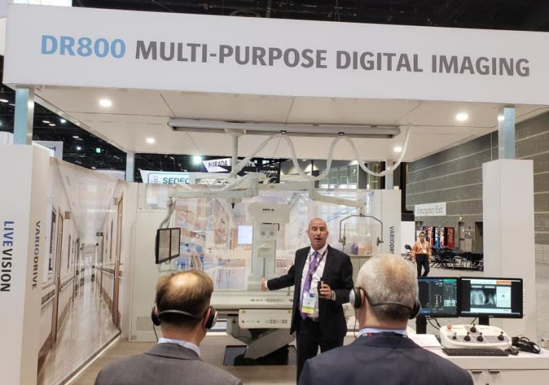
AI was arguably the most palpable element of the broader trend of efficiency in imaging, a reflection of the community-wide recognition of the need to improve processes, techniques and technologies. Exemplifying this was the DR800 from Agfa Healthcare, which appeared at RSNA 2017 as a work-in-progress (WIP) and returned to the RSNA exhibit floor in 2018 as an FDA-cleared product.
The all-purpose digital system can do both static and tilting DR exams, as well as fluoroscopic ones, including arthrography and myelography. It is designed to fit the needs of any imaging provider, according to George Curley, Agfa’s director of marketing communications. The DR800 consistently minimizes dose while delivering high-quality images thanks to Agfa’s latest generation MUSICA (Multi-Scale Image Contrast Amplification) image processing software.
Philips Healthcare sought to boost efficiency in digital radiography (DR) with its DigitalDiagnost C90. The ceiling suspended DR, which at RSNA 2018 was pending FDA clearance but available in the European Union, offers a touchscreen on its tube head and included a camera to support patient positioning.
Siemens Multix Impact uses a graphical organ program selection feature to assist techs in setting up procedures. Similarly, an onboard camera is intended to boost efficiency as it supports the adjustment of patient positioning from the control room. The company will target sales of the floor-mounted DR, which at RSNA 2018 was pending 510(k) FDA review, on community and small-size hospitals as well as clinics.
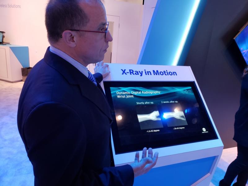
Konica Minolta redefined the term “dynamic” with a work-in-progress called Dynamic Digital Radiography (DDR). DDR fires bursts of low-dose X-rays that are recorded by digital detectors and assembled into moving images of structures inside the patient to visualize moving structures such as the lungs and joints. Fluoroscopy (which by definition is dynamic) is characterized by higher dose applications involved in examining other parts of the body and for interventional work, according to David Widmann, president and CEO of Konica Minolta Healthcare Americas. Widmann said DDR, which remains an experimental technology, has the potential to “open up an entirely new clinical space.”
Making Modalities Smart Through Deep Learning
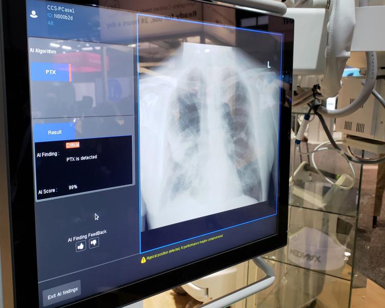
While progress in DR did not require AI, the presence of smart algorithms could be seen in multiple modalities. Exemplary of that was the Optima XR240amx, a portable X-ray machine into which GE Healthcare embedded a work-in-progress AI app for spotting collapsed lungs. The app, which is pending FDA clearance, is designed to flag chest radiographs that indicate such a pneumothorax.
This app is part of GE’s Critical Care Suite of smart algorithms and was built using GE’s newly christened Edison platform, which is meant to help accelerate the development and adoption of new technologies. GE’s clinical partners might use the newly branded platform to develop algorithms, particularly ones in AI, according to the company. The hope is that at least some of these providers will use Edison in collaboration with GE to bring advances in data processing to new applications and smart devices that the company can eventually develop into commercial products. GE and the University of California in San Francisco did just that in their development of the pneumothorax app.
Further seeking to quicken scan interpretation, GE Healthcare’s FDA-pending AIRx automates the workflow associated with brain magnetic resonance (MR). The algorithm is intended to increase consistency and productivity by automatically streamlining the choice of brain slices in scans so as to help reduce manual effort and reduce or eliminate redundancies. The idea is to make brain MR scans less dependent on the skills of technologists, thereby reducing the number of recalls due to incorrect slice placement.
Philips showcased a fledgling camera system that optically detects patient motion and, using AI, adjusts MR imaging protocols to suit, for example, variations in heart rate and respiration. “It changes the protocol and triggers the acquisitions so that all the motion related artifacts are out of the process,” said Sham Sokka, vice president and head of radiology solutions at Philips Healthcare.
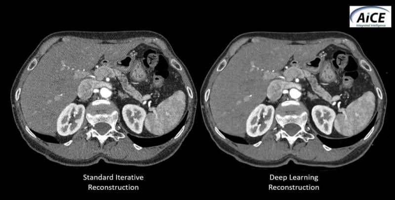
Canon showcased a smart algorithm, called the Advanced Intelligent Clear-IQ Engine (AiCE), for its ability to increase CT image resolution while minimizing patient radiation. Using deep learning technology to tell the difference between noise and signal, the AiCE algorithm “learns” to improve the resolution of CT images. It reconstructs CT images from the high-quality images produced using model-based iterative reconstruction (MBIR). Although experience with the new algorithm is limited, the company has stated that AiCE has the potential to set a new standard for image reconstruction in CT.
Leveraging AI, Siemens’ AI-Rad Companion Chest CT identifies and analyzes thoracic structures seen on CT. Siemens presented the cloud-based smart algorithm to booth visitors as a WIP pending FDA clearance. The algorithm promises to speed the interpretation of chest images by automatically measuring suspected abnormalities, such as those in the lungs; preparing reports; and guiding comparisons to prior studies. The algorithm is fully integrated into the image interpretation workflow, according to Siemens.
Preliminary testing has established that AI-Rad Companion Chest CT can identify lung lesions and calculate cardiovascular risk based on an analysis of coronary artery calcification on non-ECG-triggered CT images. The application may also help detect spine fractures due to osteoporosis.
Siemens’ Introduces AI Platforms
The chest algorithm was built on the AI-Rad Companion platform, one of two AI platforms discussed by Siemens at RSNA 2018. The other is AI-Pathway Companion, an AI-based clinical decision support system for use in diagnosis and therapy.
AI-Pathway Companion is being designed to aggregate patient history, test results and clinical studies — and then analyze the data. It might one day pull together different types of data and multi-modality images, as well as provide physicians with the patient’s clinical status. It might also suggest possible steps that physicians can take to manage their patients. Siemens expects the first clinical application coming from this platform to address prostate cancer.
Here is how an AI-Pathway Companion app might work. Scrolling through a patient’s previous and current care, the app might share patterns that could help physicians manage patients efficiently and effectively. Additionally, physicians might employ its user interface to discuss results with patients.
At RSNA 2018, Siemens also featured its syngo.Breast Care mammography reading and reporting workstation, which — by leveraging AI — is being developed to help physicians reading 2-D and breast tomosynthesis images. The WIP is being groomed to sort cases automatically and generate a score that indicates the likelihood of malignancy.
Another of Siemens’ efficiency aids, syngo Virtual Cockpit, is designed to enable remote connection to MRI, CT, PET/CT and PET/MRI scanners. As shown at RSNA 2018, it is designed to address logistical problems, improve standardization and support training, particularly regarding sophisticated or complex exams.
Other Enablers in the Radiology AI Market
Siemens Healthineers launched three MRI scanners at RSNA 2018, each based on BioMatrix technology. The BioMatrix technology, which Siemens launched in 2017, uses sensors built into the patient table to tune MR scans automatically. One patient parameter, for example, might be respiration.
BioMatrix provided the basis for the 1.5T Magnetom Altea and the 3.0T Magnetom Lumina, which during RSNA 2018 was pending FDA clearance. In addition to the advantages coming from BioMatrix, both scanners are “cost-efficient,” according to the company, and feature 70 cm bores.
Philips showcased its Ingenia Ambition X, which was announced two months earlier. The company highlighted the fully digital scanner for its negligible use of helium and workflow innovations that boost productivity.
In its first RSNA since merging with Toshiba America, Canon showed a premium 1.5T scanner. The Vantage Orian, which is pending FDA clearance, was introduced at the 2018 European Congress of Radiology in March. The company drew attention to efficient workflow built into both Orian and its 3.0T system, the Vantage Galan, which was also shown in the Canon booth.
In CT, Hitachi Healthcare Americas unveiled its Scenaria View, a premium CT that can be configured with either 64 or 128 slices. The View offers enhanced dose reduction capabilities; its 80 cm aperture and 84 kW generator accommodate large patients. A lateral shift table with a 10 cm shift range assists in patient positioning.
GE Healthcare showcased its Apex CT scanner as a WIP. The new imaging chain onboard the Apex offers improved spatial resolution and low-contrast detectability, according to the company. Images are less grainy than others generated at low radiation doses. The scanner can be equipped with a reconstruction package that the company says improves the ability to detect low-contrast structures and reduces patient radiation dose and artifacts; an image quality package that offers high image quality despite the presence of metal implants; and packages optimized for cardiac, brain and oncological scans.
AI Dominates the RSNA Expo Floor
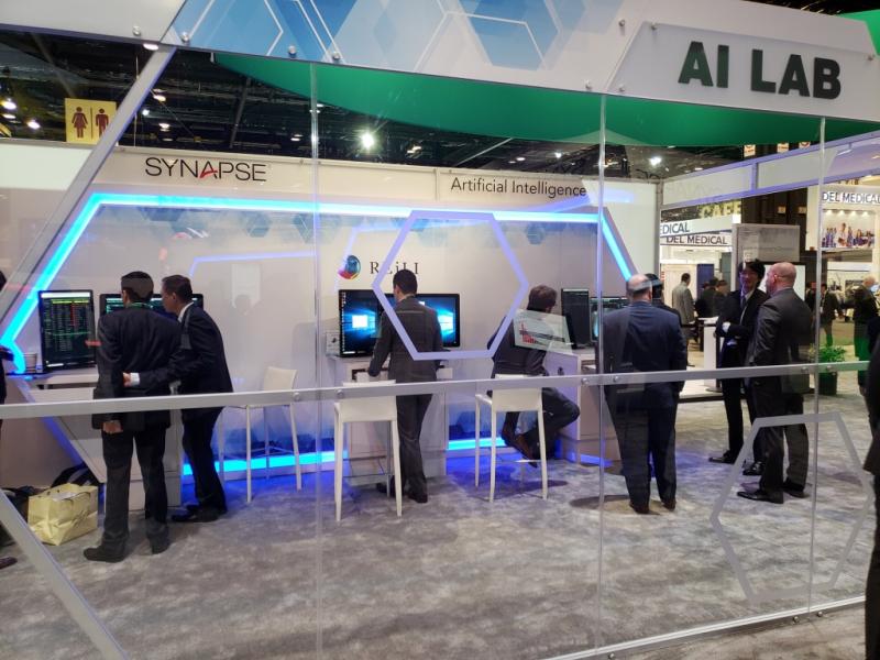
AI set the tone for advances in efficiency. The AI Laboratory in the Fujifilm Medical Systems booth presented use cases coming out of the company’s in-house AI initiative REiLI, which was unveiled at RSNA 2017 and expanded further in 2018. The AI Lab operating in the Fujifilm booth showed third-party apps, including ones by Lunit, Riverain Technologies and Koios Medical. All ran on Fujifilm’s Synapse 5, a server-side PACS, so-named because the server processes data, then transmits the results for display on workstations or other devices. Among the use cases shown were region recognition, which uses AI to recognize and extract organ regions in images, and computer-aided detection, which points out signs of pathology so as to reduce the time radiologists spend interpreting images.
The Synapse PACS, according to Bill Lacy, Fujifilm vice president of medical informatics, “serves as a bridge” between AI imaging analytics and other AI applications, one that promotes AI development among software engineers in- and outside Fujifilm.
Philips Healthcare unveiled two new twists — IntelliSpace Discovery 3.0, an advanced visualization and analysis platform designed specifically to support imaging research, and an extended version of its IntelliSpace Enterprise Edition that includes Philips’ PerformanceBridge, which optimizes site operations and workflow, according to the company.
Discovery 3.0 supports the use of clinical data to train and validate “learning” algorithms. The latest version of the Discovery platform aggregates data, normalizes them so they can be compared regardless of the make or model of equipment on which they were acquired and strips patient identifiers from the clinical data.
“It gives you the tools to create your own intelligence. It gives you the data lakes to apply it to. It gives you the tools to give validation for your algorithms,” said Jeroen Tas, chief innovation and strategy officer at Royal Philips. Providers at dozens of institutions are already using this version of the Discovery platform to develop AI algorithms.
Whereas this vendor neutral platform is designed only for researchers, Philips’ latest IntelliSpace Enterprise Edition is oriented toward routine functions. It is designed to provide access to performance data that profiles the day-to-day operation of a radiology department, according to the company. The newly refined product offers up this information as part of an interactive dashboard through the newly integrated PerformanceBridge.
Change Healthcare took a three-part approach to enterprise imaging, embodied by its own products (particularly a universal viewer and workflow intelligence products), third-party software and the in-house expertise to make them work together. The key to successful enterprise imaging is to identify goals and map a route to achieving those goals, said Todd Johnson, executive director of enterprise portfolio transformation at Change Healthcare.
“It really starts with a conversation and really listening to our customers closely,” Johnson said.
Enterprise imaging is successful when the technologies are chosen and applied in the context of carefully drawn plans, said Kim Garriott, principal consultant at the IT firm Logicalis. “Vendors can be a really big partner and a big help when organizations are looking into enterprise imaging strategies and what they should start with first,” Garriott said.
Holding the Pieces of Electromnic Medical Records Together With Artificial Intelligence
Change Healthcare exec Johnson described the company’s universal viewer, shown for the first time at RSNA 2018, as the glue that cements the myriad components of enterprise imaging, and Change’s workflow intelligence product line as the engine that orchestrates workflow.
GE showcased its Centricity Universal Viewer, noting that the viewer now supports CT, MRI and digital breast tomosynthesis, as well as patient reports such as clinical and pathology reports. Viewer updates have enhanced access to patient priors, connectivity with EMR software and exam prioritization built on iCAD’s AI technology for scoring tomosynthesis images.
Demonstrations of its vendor neutral archive, called Centricity Clinical Archive, embedded analytics for enterprise-wide IT. Its onboard Media Manager application was shown with updates that included new multimedia image capture and documentation for both mobile and desktop devices. Also debuting in the GE booth was Imaging Protocol Manager, a cloud-based imaging protocol management service for imaging modalities. Its use ensures that all imaging devices are using the same protocols, according to GE.
A lingering problem for enterprise imaging is the fragile connection between radiology and pathology, exemplified by a less than solid process for handling breast biopsies. Many radiology sites do not properly preserve biopsied tissue before it is sent to pathology.
Through its Mammotome corporate entity, Leica Biosystems proposed a solution — a specimen containment and transportation system. This system, called MammoPort, eliminates much of the handling of biopsied tissue by radiology technologists. Released commercially in September, the new product overcomes a shortcoming of breast biopsies in which biopsied tissue may be put in jars of the preservative formalin with little consideration given to the integrity of the tissue.
MammoPort could bring pathology one step closer to enterprise-wide imaging, as it tightens the connection between radiology and pathology, and helps standardize the process of transferring breast biopsy tissue from the radiology suite to the pathology lab. The tissue is placed after needle extraction into tiny trays that can be X-rayed, placed in a plastic box that is then closed, immersed in formalin and sent to pathology. This standardized tissue transfer from radiology to pathology promises to ensure quality and reduce errors.
Artificial Intelligence in Medical Imaging Builds Mojo
Among the AI algorithms shown at RSNA 2018 with a clinical perspective was the FDA-cleared IcoBrain from Belgium’s IcoMetrix, an algorithm that analyzes CT scans using deep learning (DL) to characterize traumatic brain injury. The product uses DL to quantify the severity of such typically qualitative indicators of brain injury as hyperdense volumes, compression of the basal cisterns and midline brain shift.
Leveraging DL to improve efficiency, Imalogix described how its AI algorithms can help providers comply with accreditation criteria and then help them continue to improve processes. The ultimate benefactors of these efforts is the patient, said Imalogix chairman and CEO John Heil.
If a CT scan is “off center,” for example, patients may be exposed to as much as 50 percent more radiation than necessary, Heil said. The company’s AI software reveals whether “centering” needs to be changed and how to change it, as well as how sites might get more from their installed equipment.
One Imalogix product is being sold by contrast vendor Guerbet to track patient exposure to ionizing radiation. The French company, best known for contrast media, featured an Imalogix-built product alongside its own Contrast&Care software, which is designed to improve the productivity and traceability of Guerbet contrast media.
Guerbet focused heavily on digital offerings at RSNA 2018, also showing an AI tool developed by IBM Watson Health, which summarizes patient information into single-view reports. The company is working jointly with IBM Watson Health to develop AI software to support liver cancer diagnosis and care as part of its GEAR 2023 plan, Guerbet’s five-year strategic plan, which heavily stresses increased digitalization.
“We will probably develop other applications in the same space,” said David Hale, chief commercial officer for diagnostic imaging at Guerbet, “but always where we think we can provide diagnostic confidence help and productivity help for our customers.”
At RSNA 2018, iCAD unveiled its ProFound AI, which leverages AI to detect cancer in breast tomosynthesis. The software is geared toward screening, but can also be used for diagnostic studies. It examines every image in the tomosynthesis scan, detecting malignant soft tissue densities and calcifications.
The algorithm calculates “Certainty of Finding” to each detection and “Case Scores” for each case to represent the algorithm’s confidence that a detection or case is malignant. The scores are represented on a scale from 0 to 100 percent. High scores indicate high levels of confidence in the malignancy of the detection or case, explained Rodney Hawkins, vice president of marketing at iCAD. As such, they serve as guides to aid in determining if suspicious findings or cases need further study. The use of ProFound AI could lead to improved detection, fewer patient recalls and mammographers spending less time reading images, Hawkins said. “Because of the time-saving aspect, it is really geared toward a screening environment. But it can be used for both,” he explained.
From its booth within the Machine Learning Showcase, Arterys promoted its recently FDA-cleared Cardio AIMR, which analyzes MR images for cardiovascular blood flow. The company also showed AI-based tools that measure and track liver lesions and lung nodules; accelerate the display and reading of medical images; and support the display of mammograms using the Google Chrome web browser.
While AI was the most obvious trend at RSNA 2018, its presence hinged on the need to improve efficiency. Enroute to that, radiologists need to be watchful.
“AI can fail. And often there are patterns to the way it can fail,” Erickson said. “We need to learn those and address those and continually improve the algorithms.”
Greg Freiherr has reported on developments in radiology since 1983. He runs the consulting service, The Freiherr Group.
Related RSNA AI Coverage:
VIDEO: Editor’s Choice of the Most Innovative New Artificial Intelligence Technologies at RSNA 2018
VIDEO: Technology Report: Artificial Intelligence
How to Market Healthcare Artificial Intelligence Software
Selecting an AI Marketplace for Radiology: Key Considerations for Healthcare Providers

 April 23, 2024


