Image courtesy of Cerner
Mammography has been the gold standard for breast cancer screening ever since it was proven to reduce mortality in clinical trials in the 1960s. A 2015 report from the U.S. Department of Health and Human Services (HHS) revealed, however, that only 66.8 percent of women age 40 and over — the demographic traditionally targeted for screening — had received a mammogram in the previous two years.1 Understanding of fibroglandular breast density and its impact on cancer detection, as well as new screening guidelines that challenge conventional wisdom, are among the factors impacting compliance rates, and they suggest that screening has evolved beyond the one-size-fits-all approach of using mammography alone.
Gerald Kolb, JD, president of The Breast Group in Bend, Ore., shared this sentiment in a presentation to radiology administrators at the 2016 annual meeting of the Association for Medical Imaging Management (AHRA) in Nashville. He argued that switching to a more personalized, multimodality approach of screening would help improve both compliance and cancer detection rates, which in turn will help keep the cost of care down.
A Brief Overview of Screening
To fully understand the necessity of multimodality screening, a brief look at how breast cancer screening has evolved is necessary.
Mammography. Following the establishment of mammography in the 1960s, screening continued to expand for the next decade. The first paper was released in 1976 discussing the impact of parenchymal density patterns on mammography, indicating that alternative screening methods may be required to ensure the highest probability of detecting early-stage cancers.
Ultrasound. Ultrasound was one of the first alternative modalities explored, with the first paper released in 1995 showing its propensity for detecting mammographically occult cancers. A series of studies followed, underscoring the potential of ultrasound as a supplemental screening option for women with dense fibroglandular breast tissue. Most recently, a 2015 study in the Journal of the National Cancer Institute (JNCI) compared cancer detection rates between ultrasound and mammography in 2,809 patients across the United States, Canada and Argentina. Results showed a larger proportion of invasive and node-negative cancers in patients who also underwent ultrasound, with overall detection rates comparable to mammography despite more false-positives.2
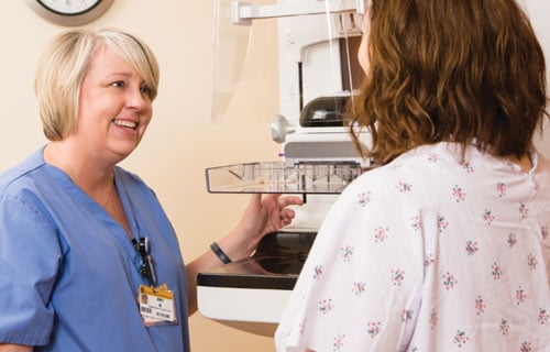
Full-field Digital Mammography. Full-field digital mammography (FFDM) followed breast ultrasound in 2000 when GE Healthcare gained U.S. Food and Drug Administration (FDA) approval for the first FFDM unit.
Breast MRI. Breast magnetic resonance imaging (MRI) became part of screening recommendations for the first time in 2007 through the American Cancer Society, and those guidelines are still in use today. They recommend MRI as supplemental imaging for women at the highest risk of breast cancer (high risk is defined as a 20-25 percent lifetime risk under the BRCA PRO and other mathematical risk models).
Digital Breast Tomosynthesis. Hologic brought digital breast tomosynthesis (DBT), also known as 3-D mammography, to the market following FDA approval in 2011. DBT acquires breast images in multiple 3-D slices, offering a much clearer picture than traditional 2-D mammography. This helps improve cancer detection rates while simultaneously reducing recalls.
New Insurance Guidelines Offer Mixed Messages
Patient and provider decisions on which screening modalities to employ are largely based on insurance
coverage, which since the passage of the Affordable Care Act, is shepherded by the U.S. Preventive Services Task Force (USPSTF). The task force released new guidelines for breast screening earlier this year, which caused heated debate in the radiology community. Most significantly, the task force gave a lower grade recommendation for annual mammography for women in their 40s, saying the benefits may not outweigh the potential risks. The lower grade means that insurance providers are no longer required to cover the procedure for that age group — a deeply troubling reversal for many in the radiology community because, as Kolb noted, “half of the life-years lost to breast cancer are lost in that group.” The task force also reduced the recommended frequency of mammography to every other year for women 50-74.
While insurers may be shifting their views on traditional mammography, 3-D mammography took a major step toward acceptance when Cigna announced in August it would become the first national insurer to cover the technology, as of Aug. 23. The company said that it changed its policy when the USPSTF guidelines were released, and based the new coverage off of more recent guidelines from the National Comprehensive Cancer Network (NCCN).
Breast Density Inform Laws
As technology has continued to advance, the conversation about breast density has moved into the national spotlight, with patients demanding full disclosure so as to make the most informed decision about their own care. The groundswell reached the legislative level in 2009, when Connecticut became the first state to pass a breast density inform law. In the intervening seven years, 27 other states have added similar legislation. Each law is worded differently and has slightly different provisions (some require all patients to be notified about density, while others only require notification if the patient is determined to have dense breasts), but the same spirit of open communication pervades across the board.
Implementation and Education
Even if a facility has weighed these mitigating factors and decided the benefits of multimodality screening outweigh the challenges, implementing such a strategy has its own set of challenges. In addition, the practice must consider how best to educate its patients about newer technologies and concepts so they can make informed decisions as part of their own healthcare team.
Regardless of what technologies are available, Kolb said that a mammogram should still always be the first step in the screening process. It is from this initial mammogram that the radiologist can determine if supplemental screening is needed based on the woman’s fibroglandular density. Several vendors offer breast density analysis software. These programs offer different methods for objectively calculating the ratio of dense to fatty tissue.
During this stage, breast density and its relevance should be explained to the patient, who may have heard of the concept but will likely have questions. The notification required by most density inform laws tells patients to speak to their primary care physician — a piece of advice Kolb thinks may be misguided.
“Breast density is a radiologic phenomenon. The density is on the image, not in the tissue itself,” he said. “[Physicians] don’t typically have any training at all regarding breast density and its implications.” In his view, the radiologic technologist is in the ideal educational position, as they have the most direct contact with the patient.
Technologists at The Breast Group play an active role in the education process, which intersects with the patient care continuum at several points. Initially, a brochure about density is mailed to the patient along with their reminder letter when a scheduled mammogram is approaching. In case this step was missed, the same brochure is distributed to each patient at registration on-site.
Kolb has his technologists initiate the density discussion with patients while the exam is ongoing — specifically after the fourth view is acquired. All technologists have a script to follow as well as visual aids, and Kolb estimates the entire process takes about 90 seconds. “That actually is very supportive of the referring physicians, because radiology has provided a basis for the discussion and decision-making that may occur later,” he said. It is only after this conversation, when the patient has a full understanding of breast density and its implications, that the technologist shares their individual, objective density and discusses supplemental imaging.
If supplemental imaging is required, study data suggests that performing the second exam the same day is the best way to ensure compliance.
Opening the Lines of Communication
Kolb stressed that communication between the mammography facility and the referring physician is critical to practicing patient-centered, coordinated care. The physician must fully understand the benefits of early cancer detection and how breast imaging can help.
The Breast Group recommends sending a personal letter and breast density memo to each referring physician in your area to get the ball rolling. For your top 10 referrers, follow-up personal contact is highly recommended.
Another way to help streamline the breast imaging process is to have the referring physician write a conditional contingent order for the screening. In radiology, these documents initially request a mammogram, but immediately authorize further imaging if it is required — i.e., if the patient is determined to have dense breasts.
Related Breast Imaging Technology Articles:
Implementing Advanced Breast Imaging Technology
Recent Trends and Developments in Contrast Media
References

 April 18, 2024


