Breast imaging technologies have seen a rapid evolution.
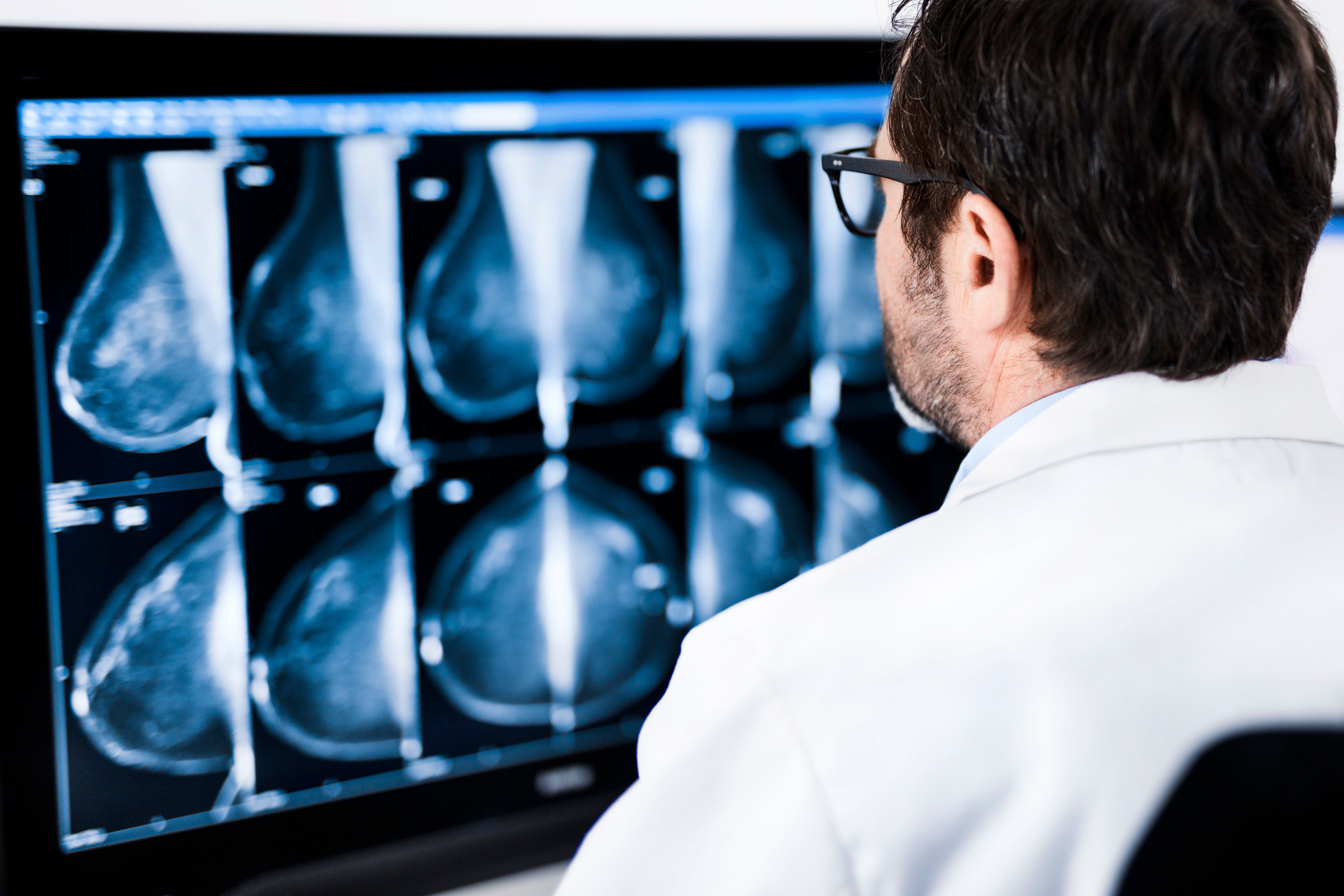
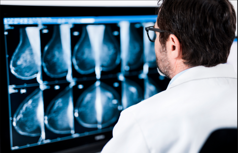
Breast imaging technologies have evolved rapidly in the last two decades to help physicians detect breast cancers at an earlier stage. Clinical studies have proved the effectiveness of newer technologies like automated breast ultrasound (ABUS), digital breast tomosynthesis (DBT) and breast magnetic resonance imaging (MRI) in finding more cancers that traditional mammograms might miss as well as interval cancers that develop between screenings.
While the benefits of these and other supplemental screening methods have been demonstrated, a multimodality approach is not necessarily the best one for every woman. “It’s not a one-size-fits-all, it never has been in a lot of ways,” said Karen Hou, M.D., diagnostic radiologist at Northwestern Medicine Central DuPage Hospital (CDH) in Winfield, Ill. To ensure that every woman gets the appropriate level of care she deserves, Hou and her colleagues at the CDH Breast Health Center opened the CDH High-Risk Breast Clinic in 2017.
Benefits vs. Harms of Breast Cancer Screening
The frequency of mammography screening has become a contentious topic in recent years, largely due to the most recent updates to screening recommendations from the U.S. Preventive Services Task Force (USPSTF), an independent, volunteer panel of experts in evidence-based medicine. In 2009, the Task Force recommended against routine mammography screening for women ages 40-49 with a grade of “C”, a controversial stance among healthcare providers. Instead, the Task Force stated that women under age 50 should decide with their doctor whether biennial screening would be appropriate. The USPSTF also recommended biennial screening mammograms for women between the ages of 50 and 74, with a “B” recommendation.1 The task force finalized these recommendations in 2016.
USPSTF grades are based on a review of the indicated preventive service, balancing the harms and benefits of the service. One of the harms most often associated with breast cancer screening is the risk of overdiagnosis. A 2017 study out of Denmark found that breast cancer screening in that country was associated with a substantial increase in the incidence of nonadvanced tumors and ductal carcinoma in situ (DCIS), but not with a reduction in the incidence of advanced tumors. The researchers concluded that “it is likely that one in every three invasive tumors and cases of DCIS diagnosed in women offered screening represent overdiagnosis,” an incidence increase of 48.3 percent.2 An American study published last June in the New England Journal of Medicine reached similar conclusions, finding that screening tended to overdiagnose small, slow-growing tumors, particularly in older patients.3
Hou also noted that many women are fearful of the radiation associated with mammograms. “But truly with mammography there’s very little risk,” said Hou. “It’s overblown in a lot of people’s minds. But again, it’s something we want to be mindful of.”
Individualizing Breast Cancer Screening
Taking this approach of weighing benefits against harms, the High-Risk Clinic — which operates at CDH and nearby Northwestern Medicine Delnor Hospital — is designed to identify individuals at higher risk of breast cancer who may benefit from additional screening.
The clinic’s website indicates that high-risk assessment is reserved for women who meet any of the following criteria:
• Multiple family members with breast and/or ovarian cancer;
• First-degree relative (mother, father, sister, daughter) with a diagnosis of breast and/or ovarian cancer;
• Breast biopsy with pathology showing atypical cells or lobular carcinoma in situ;
• Have a known genetic mutation that increases the risk of developing breast cancer; and/or
• History of prior chest radiation therapy.
Mammography is still the standard of care at Northwestern Medicine CDH Breast Health Center, which is designated an American College of Radiology (ACR) Breast Imaging Center of Excellence and has been recognized through the American College of Surgeons’ National Accreditation Program for Breast Centers (NAPBC). Mammography exams are conducted during a 15-minute appointment. Hou said these exams are typically read within a few days, and that “most women will have a normal screening exam.” If the exam is abnormal in any way, the hospital will reach out to the patient to come back for a diagnostic workup. Diagnostic exams at the hospital consist of a follow-up mammogram concurrent with ultrasound imaging. If necessary, the hospital also offers on-site biopsy services.
If deemed appropriate, the team at the High-Risk Breast Clinic will make arrangements for genetic testing, medications and consultation regarding prophylactic surgery. They will also coordinate with the patient’s primary care provider throughout the process to ensure smooth transitions and compliance with medical recommendations. Patients deemed moderate- to high-risk are offered ongoing management and surveillance at the clinic. This includes services like education on proper breast checks and modifiable risk factors such as diet, exercise, tobacco and alcohol use. The clinic also offers social work and emotional support services to help patients and their families handle the realities of cancer care. “They can sit down with somebody to really discuss, ‘Am I at risk and what should I be doing?’ ” said Hou.
A year into the endeavor, she believes the clinic has helped find and treat more cancers, but perhaps more importantly, helped patients better understand and take control of their health.
Breast Density and Patient Education
Hou noted that patients are coming in with more knowledge of their health — or at least more questions — than in the past. Many of their questions revolve around fibroglandular breast density, a cancer risk factor that has come to the forefront of women’s health conversations in recent years.
If a woman has dense fibroglandular breast tissue it can obscure tumors on a 2-D mammogram, as the tumor and the dense tissue both appear white on the image. Prior to 2009, facilities were not required to disclose information about breast density to patients, and the consensus among providers was that telling patients would do more harm than good by increasing anxiety. In 2009, states started passing individual breast density inform laws requiring some level of information in a woman’s mammography report if she was found to have dense breast tissue. Today, 34 states have density inform laws on the books. The increased awareness of issues like fibroglandular breast density has augmented the importance of supplemental modalities like ABUS and DBT.
Illinois does not have a breast density inform law (although bills were introduced in the state House and Senate in February 2018), but patient education is still a priority at Northwestern Medicine CDH. Hou said the hospital does inform women in their mammography reports if they have dense tissue and could benefit from supplemental screening. They have also worked to ensure that their network of referring physicians and primary care providers are knowledgeable about the options available to their patients. “We really try to help patients understand in the venues that we’re able to, and their doctors, about understanding that [dense breast tissue is] not a bad thing, it’s just the way it is,” Hou said. She added that consistency is an important consideration as well — they wanted to have someplace that referring physicians could send their patients and ensure that patients would have their own specialist that they could see regularly.
Implementing Advanced Imaging Technologies
While Hou touted the benefits of newer technologies like ABUS and tomosynthesis, she did acknowledge that they come with a learning curve — especially ABUS. “The first six months or so are the hardest when the technologists are trying to get comfortable with doing it and we are constantly critiquing them,” she said. “We would check all of their work in the beginning, and auditing is really important for us because it’s feedback for us to learn what we’re doing.”
According to Hou, the key to integrating new breast imaging technologies is figuring out how to work them in while minimizing disruptions. “You have to look at how you want it to fit into your workflow. You have to make a decision ‘I’m going to use this as a screening tool,’ ” she said.
Ultimately, the decision should come down to what is best for the patient. “We want to be applying the best technology in the most appropriate way that will benefit people the most. What we’ve done is try to use some logic along with what is most accessible for people and what we could do to best serve them,” Hou concluded.
Recent Related Breast Imaging Content:
VIDEO: The Impact of Breast Density Technology and Legislation
Greater Evidence, Payment Expansion Driving Tomosynthesis Adoption
Technology to Watch in Breast Imaging
The Transition to 3-D Breast Imaging
References
1. U.S. Preventive Services Task Force. Screening for Breast Cancer: U.S. Preventive Services Task Force Recommendation Statement. Annals of Internal Medicine. Nov. 17, 2009.
2. Jørgensen K.J., Gøtzsche P.C., Kalager M., et al. Breast Cancer Screening in Denmark: A Cohort Study of Tumor Size and Overdiagnosis. Annals of Internal Medicine, March 7, 2017. http://annals.org/aim/article-abstract/2596394/breast-cancer-screening-denmark-cohort-study-tumor-size-overdiagnosis
3. Lannin D.R., Wang S. Are Small Breast Cancers Good because They Are Small or Small because They Are Good? New England Journal of Medicine, June 8, 2017. https://www.nejm.org/doi/full/10.1056/NEJMsr1613680

 April 18, 2024


