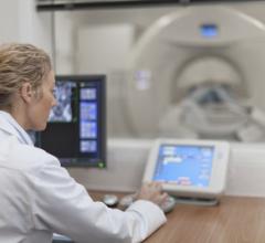
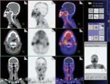
GE Healthcare's Discovery STE clinical imaging assists with diagnosis.
Image fusion — combining image data from different modalities, and of which hybrid imaging is a subset — is revolutionizing the way physicians view and treat disease. The process can be performed through computer workstations and software, however, dedicated hybrid systems, such as PET/CT and more recently, SPECT/CT, minimize the drawbacks of fusing images derived from two separate pieces of equipment. According to some experts, dedicated hybrid systems possess the potential to bring the diagnosis and treatment of cancer, neurological and cardiac diseases to the brink of transformation. These hybrid units, which allow the patient to remain on the same exam table for both scans, produce almost perfectly aligned images, leading to earlier, more accurate detection and treatment and, ultimately, better patient outcomes.
First Line of Defense
Since its introduction to the medical imaging market over 30 years ago, CT has been a diagnostic favorite because of its ability to present anatomical information in precise detail. In recent years, CT has graduated from producing single slices to generating eight, 16 and even 64 slices of data in one breathhold.
By 1994, when PET physicist David Townsend, Ph.D. and Ronald Nutt, Ph.D. received a grant from the National Institutes of Health (NIH) to build the first PET/CT prototype, PET was already recognized as an important diagnostic tool. Today, based on Drs. Townsend and Nutt’s collaboration, the ability to combine physiological data from a PET exam with CT’s anatomical image slices has made PET/CT the number one imaging tool in oncology, and its potential for other clinical applications is growing. Over 1,000 PET/CT systems are currently in clinical use, said Dr. Townsend. Results of PET/CT scans not only enhance cancer detection, but also aid in the staging, treatment and follow-up of the disease, allowing physicians to personalize treatment plans.
“As data demonstrating the advantages of PET/CT over magnetic resonance and CT images in early disease detection continue to accumulate, it is reasonable to anticipate that PET/CT imaging systems could become the ‘first line of defense’ in optimization of treatment for cancer patients,” according to Jacqueline Brunetti, M.D., director of Radiology, Holy Name Hospital, Morristown, NJ.
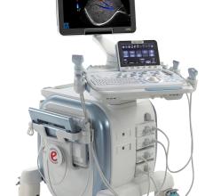
The first to utilize the Discovery STE, a PET/CT platform from GE Healthcare, for neurological applications is Satoshi Minoshima, M.D., professor and vice chair for Research at the University of Washington Department of Radiology.
“PET/CT is a great diagnostic tool and powerful enough to accurately and quickly image brain metabolic abnormalities associated with neurological disorders such as Alzheimer’s diseases,” said Dr. Minoshima. “Discovery STE delivers superb quality of brain images.”
The new Discovery VCT, according to GE, is the world’s first true 64-slice combination PET and volume CT system for cardiac imaging applications, providing clinicians with the diagnostic confidence necessary for assessing coronary artery disease.
Southwoods X-ray and Open MRI in Boardman, OH became the first beta site for the Hitachi SceptreP3 PET/CT in late 2004, and by January 2005, the system was completely operational. Since that time, says Dr. Steven Aubul, medical director at the facility, the group’s two technologists have been able to scan patients quicker, while the radiologists enjoy the increased accuracy. “Having the anatomic information and the metabolic information allows you to narrow down whether something is a real finding or not, and if it is a real finding, exactly where it is in the body,” he said. “That can make a big difference in how something is treated or how something is biopsied.”
SceptreP3’s Dual Attenuation Correction (DAC) technology allows the combination of both CT and sealed source attenuation correction to effectively image patients with metal implants. In addition, the Non-Rigid Fusion7-D algorithm provides precise registration by correcting for respiration differences between PET and CT acquisitions, and the integration of the AVIA-PACS architecture improves software communication, multimodal connectivity and supports RAID archiving capabilities.
The facility currently uses the SceptreP3 for oncologic imaging only, but Dr. Aubul said they are investigating cardiac applications. All in all, Southwoods X-ray and Open MRI is quite satisfied with the system and its workstation, explaining it produces high-quality images. What Dr. Aubul does see a need for, however, is more patients, explaining that referring physicians still require a lot of education as to the applications and benefits of PET/CT.
Improving the Diagnostic Process
SPECT is most commonly used for whole-body bone imaging, cardiac imaging and brain-perfusion studies. According to Homer A. Macapinlac, M.D., associate professor of Radiology, M.D. Anderson Cancer Center, Houston, TX, ”SPECT and CT are each very powerful modalities, and together there is synergy and the possibility to improve diagnosis and therapy for the patient. The introduction of this new technology offers the chance to re-examine how the diagnostic process works — the order in which studies are performed and how the whole care pathway is constructed.”
In a single scanning session, the imaging technology of Siemens’ Symbia TruePoint SPECT/CT quickly captures comprehensive, accurate information on both the molecular and anatomical levels, enabling clinicians to detect changes in molecular activity even before structural changes become visible. Nuclear medicine professionals at the University of Michigan Health System are experiencing these benefits first hand. James R. Corbett, M.D., professor of Radiology and Internal Medicine, expects TruePoint SPECT/CT technology to play an important role in the continued development of molecular imaging capabilities for SPECT. “[TruePoint SPECT/CT] will probably change the way we do everything within the next few years,” he explained, as it facilitates diagnosis and molecular and radiographic therapy.
Launched at the Society of Nuclear Medicine (SNM) meeting in June 2004, the Precedence SPECT/CT from Philips features unique imaging algorithms combined with 16-slice CT to facilitate myocardial perfusion and coronary artery imaging. The solution provides registered and individual SPECT and CT as well as attenuation-corrected nuclear medicine images.
Not for Everyone?
Adding to hybrid imaging technology’s popularity is the increased coverage by CMS for PET/CT. According to a Frost & Sullivan research report published Sept. 30, 2005, SPECT/CT has the potential to attract the same reimbursement levels as PET.
Despite increased reimbursement, not to mention the documented clinical and workflow benefits, skeptics may question whether dedicated hybrid scanners are really necessary. Can’t the same end result, combining physiological data with anatomical data to produce a more accurate image of what is happening inside the body, be accomplished through fusion software, without having to invest in dedicated hybrid systems?
Much, of course, may depend on the size of the facility, the volume of studies they perform as well as the type of studies. According to Dr. Townsend, professor of Medicine and director of Cancer Imaging and the Tracer Development Research Program at the University of Tennessee Medical Center, fusion software is not an effective solution for imaging outside the brain because of internal organs’ tendency to shift during or between scans. He added that logistical considerations such as installation, siting and workflow can be more of a concern than cost when weighing the purchase of a dedicated hybrid scanner.
With PET/CT its favored tool, oncology is expected to further the demand for hybrid imaging, and in the process, cost justification for the technology. Now that stand-alone PET systems are nearly obsolete, there is no turning back; the future for oncology applications and more lies in hybrid imaging. Driven by 64-slice CT, cardiology applications are also poised to reap the benefits. Helping to ease the transition for physicians and technologists are PET/CT and SPECT/CT courses and hands-on workshops, such as those sponsored by the SNM and the GE-sponsored Molecular Imaging Masters Series held this year in the U.S. and Canada.
By most accounts, the consensus is clear: The fusing of form and function is propelling nuclear medicine to a new level of diagnostic and treatment accuracy.
 August 01, 2022