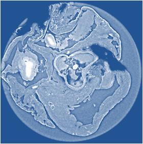
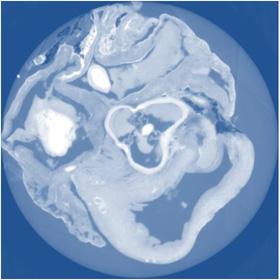
This image shows a non-invasive \"slice\" of a rat's heart tissue made using grating-based X-ray phase tomography, which excels in contrast, albeit with some blurring when compared to propagation-based phase-contrast X-ray imaging.
January 15, 2015 — X-ray phase tomography is an imaging technique that uses penetrating X-rays to create volumetric views through "slices" or sections of soft biological tissues, such as tumors, and it offers strongly enhanced contrast compared to conventional CT scans. Yet scientists still do not know which X-ray phase tomography methods are best suited to yield optimized results for a wide variety of conditions.
To answer this question, a large group of researchers in Europe compared three different X-ray phase tomography methods at the European Synchrotron Radiation Facility's (ESRF) beamline ID19 in France — X-ray grating interferometry, propagation-based phase tomography with single-distance phase reconstruction and holotomography.
Led by Irene Zanette, a scientist affiliated with both ESRF and the Technische Universität München (TUM) in Germany, the researchers examined cancerous tissue from a mouse model and an entire rat's heart, which they report in the Journal of Applied Physics, from AIP Publishing.
Along with colleagues Bert Müller, group leader of the Biomaterials Science Center in Switzerland, and Timm Weitkamp, a scientist at the Synchrotron SOLEIL in France, the team explored which method performs best in terms of spatial resolution and visualization/quantification of relevant features in the samples. They also investigated other related factors such as the simplicity of the setup, and the data acquisition and analysis involved in each method.
The researchers chose to exploit synchrotron radiation, which produces significantly higher-quality X-rays than conventional X-ray generators such as those found in hospitals.
"Think of synchrotron radiation as being analogous to the sort of monochromatic, collimated and intense light produced by lasers, while conventional X-ray generators in hospitals are more analogous to light bulbs we use within our homes," explained Zanette.
Zanette explained that while their study was performed using synchrotron radiation, the same techniques are amenable to both polychromatic and divergent beams and can also be implemented at conventional X-ray sources.
The researchers used an advanced X-ray technique known as "phase-contrast imaging." This type of imaging works by making the X-ray beam interfere while it propagates from sample to detector, according to Zanette. "This interference is fundamental because it encodes precious information on the phase of the X-ray waves."
By comparison, conventional X-ray imaging — of the sort performed at hospitals and airports — does not use phase information. Rather, it relies only on the attenuation of the amplitude (reduction in intensity) of the X-ray waves by the object under study to generate image contrast.
"More detailed information is contained in the phase than the amplitude, so it enables us to obtain images with much greater contrast and clearly differentiates cancerous tissue from healthy tissue," Zanette said.
The researchers were able to show that for each specimen, the spatial resolution derived from the characteristic morphological features is about twice as good for holotomography and single-distance phase reconstruction compared to X-ray grating interferometry. They also found that X-ray grating interferometry data generally provide much better contrast-to-noise ratios for anatomical features, excel in fidelity of the density measurements, and are more robust against low-frequency artifacts than holotomography.
The researchers regard all three of the phase tomography methods as being complementary. "The application determines which spatial and density resolutions are desired for the imaging task and dose requirements, so it really comes down to a choice between the complexity of the experimental setup and the data processing," noted Müller. "It's important to choose the ideal technique for your specific purposes."
Since synchrotron radiation is of higher quality than the radiation at conventional sources, measurements at synchrotrons represent benchmarking experiments when translating these tomography techniques to clinical practice — especially X-ray grating interferometry, which is attracting attention for use in hospitals.
The article, "Experimental comparison of grating- and propagation-based x-ray phase tomography of soft tissue," is authored by Sabrina Lang, Irene Zanette, Marco Dominietto, Max Langer, Alexander Rack, Georg Schulz, Geraldine Le Duc, Christian David, Jürgen Mohr, Franz Pfeiffer, Bert Müller and Timm Weitkamp. The authors of the paper are affiliated with Technische Universität München in Germany, Biomaterials Science Center in Switzerland, and Synchrotron SOLEIL in France.
For more information: www.jap.aip.org

 April 16, 2024