This article appeared as an introduction to the CT Simulation Comparison Chart in the October 2010 issue of Imaging Technology News.
Computed tomography (CT) virtual simulation systems allow 3-D viewing and planning along with verification images to achieve the same level of treatment simulation as with radiotherapy (RT) simulation. CT offers an improvement over conventional simulation because of its 3-D visualization, only one session is needed with the patient and volume markup can be done without the patient being present.
Introduced in the 1990s, CT simulation is now the primary modality used for RT simulation and is required for intensity-modulated radiotherapy (IMRT), which allows a high dose of radiation to be delivered to a tumor, while minimizing irradiation of surrounding healthy tissue.
CT virtual simulation makes it possible to deal with interfraction motion (i.e., changes in target position due to set-up error or naturally-occurring changes in organ position over time) and intrafraction motion (i.e., organ motion during treatment, usually due to respiration or other physiological processes).
How it Works
CT simulation requires an actual CT scan of the patient followed by virtual simulation, for which the patient is not required. These systems use a CT scanner with specialized hardware and software for RT simulation and treatment field delineation. Usually the scanner is networked to an RT treatment planning system.
The CT-based simulation delineates a tumor on the CT slices, the target volume is determined using treatment planning software and the treatment field is selected. The simulation is then used to laser mark the patient for radiation treatment. Imaging, treatment planning, treatment field delineation and patient marking is completed in a single session.
The simulations are also used to verify tumor margins and surrounding anatomy to track tumor regression and 3-D image analysis for multileaf collimators and conformal radiotherapy.
Since RT is administered with the patient on a flat couch, CT scanning is performed with a patient lying in the treatment position on a flat couch top or removable flat-pad insert. The curved tabletop used for diagnostic imaging alters the position of internal anatomy in relation to external anatomical landmarks.
The computer is used to localize the tumor, delineate the target and calculate coordinates to mark the patient’s skin with a laser. The laser mark indicates where to mark the patient’s skin at the intersection of the target volume center. Beam’s-eye-view images can be displayed and printed to visualize the planned treatment beam in relation to the anatomy.
Digitally reconstructed radiographs (DRRs) act as maps representing beam attenuation along rays drawn from the radiation source. They are comparable to simulator films and are generated at various beam orientations to verify or modify patient positioning, collimator angles, beam width, SAD (source-axis distance) and gantry positioning during treatment.
Recent Innovations
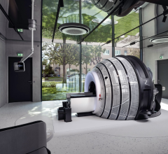
The Siemens CT Vision system combines a Somatom CT scanner on rails with a linear accelerator and treatment table. With a CT moving on rails and a smoothly rotating treatment table, it is possible to perform a CT scan and deliver treatment in a single time slot in the same room.
Resonant Medical has developed 3-D soft tissue planning, image-guidance and adaptive radiotherapy software. Its Clarity System is both noninvasive and nonionizing for image-guided radiation therapy (IGRT) for soft tissue cancers, such as prostate and breast. The Clarity System is designed to enable radiation oncologists to visualize the target anatomy during each step of the patient flow from simulation and planning through treatment.
In June, Brainlab AG signed a deal with Toshiba Medical Systems to integrate the Aquilion LB CT with the Brainlab Brainsuite iCT digitally integrated operating room. The agreement will enable fast IMRT, focused IGRT with high-precision treatment planning. Brainsuite iCT integrates intraoperative CT scans with surgical planning and ceiling-mounted navigation. Up-to-date images combined with navigation provide surgeons with immediate quality control and the ability to check their progress before concluding surgery. The 16-slice Aquilion LB features a large gantry aperture of 90 cm and a scan field of 70 cm, covering more anatomy.
The Philips Brilliance CT Big Bore platform for oncology was released in November 2009. It is designed to increase accuracy in lesion localization, improve efficiency in image reconstruction and streamline workflow across patient marking and treatment stages. An amplitude binning algorithm, through 4-D correlated imaging, allows patients to breathe freely while capturing and sorting images according to inspiration/expiration data points along a respiratory waveform. This is especially beneficial for patients that have difficulty holding their breath.

 April 17, 2024