At the surface, the term "advanced visualization" seems nebulous at best. Medical imaging technology continues to develop and advance at a rapid rate, so determining what qualifies as advanced remains a moving target as well. In general, however, the current cutting edge of imaging centers around 3-D/4-D viewing and mobile technology, with different specialties implementing them in unique ways.
3-D and 4-D
Once cumbersome and impractical, three- and four-dimensional viewing now form the cornerstone of many advanced visualization systems and are becoming more commonplace as technology evolves.
Brit Systems launched an optional 3-D add-on for its WebWorks zero-footprint viewer at the 2014 Radiological Society of North America (RSNA) annual meeting, providing a brand-new toolset for users. All 3-D renderings are fully rotatable, with measurement tools including maximum intensity projection (MIP) and multiplanar reconstruction (MPR). The system can upload DICOM and non-DICOM images, and non-DICOM images can be saved as DICOM objects. WebWorks 3-D utilizes the emerging Image Object Change Management (IOCM) standard, so any changes made in the viewer are automatically saved anywhere the image is stored.
Some manufacturers are taking a different route and building advanced visualization capabilities directly into their scanners. Carestream Health is in the process of creating a 3-D cone beam computed tomography (CBCT) system specifically for imaging orthopedic, weight-bearing extremities including the knees, legs and feet. Compared to traditional full-body CT scanners, cone beam CT lowers both radiation exposure and overall cost.
GE introduced 4-D viewing to its Voluson ultrasound scanners with the release of the Voluson E10 last October. With this advance, both the technician and patient can watch the fetus move in real time, with HDlive software enabling precise calculation of depth, shape and detail.
Siemens introduced advanced visualization functionality to its Artis zee, Artis Q and Artis Q.zen angiography systems via the new PURE platform at the 2015 American College of Cardiology (ACC) meeting. PURE adds several new features to all three systems, such as a 3-D Wizard for one-click 3-D image acquisition, with the user simply selecting the desired image type for the body part in question; QuickZoom, which centers the field of view around the desired point while rotating the image or adjusting the zoom factor on syndoDynaCT images; and 2-D/3-D Fusion, which allows fusion of any pre-procedural CT, magnetic resonance imaging (MRI) or positron emission tomography (PET) images utilizing two fluoroscopic images for live image guidance.
Advanced Visualization in Cardiology
In terms of the various specialties, cardiology has been one of the most prolific when it comes to new advanced visualization products. This upswing is largely due to several major advances in interventional solutions, including transcatheter aortic valve replacement (TAVR) devices, left atrial appendage (LAA) occluders and the MitraClip. The high degree of complexity of these procedures - along with transcather mitral valve replacement (TMVR), which has yet to fully develop in the United States - demands higher-quality images to guide device implantation.
Blood flow visualization and analysis, in particular, are crucial for proper implantation, and transesophageal echocardiography (TEE) is the imaging method most often specified. Several vendors have begun adding blood flow analysis to their solutions. Siemens introduced the eSie Valves analysis package on its Acuson SC2000 Prime cardiac ultrasound system; the software can provide semi-automated measurements of both the mitral and aortic valve within seconds.
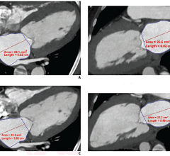
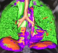
Version 7.0 of Philips' IntelliSpace Portal, introduced at RSNA 2014, features a host of advanced cardiac imaging applications, highlighted by Advanced Vessel Analysis and TAVI Planning for CT (computed tomography). The former module offers a series of tools for detailed inspection of contrast-enhanced vessels, and can even remove bony structures obscuring vascular structures with one click; the latter allows rapid assessment of the aortic root for proper device sizing.
Agfa HealthCare entered the cardiac advanced vis market at ACC.15, introducing its Enterprise Imaging for Cardiology Suite. Agfa partnered with TomTec to take advantage of the latter's multimodality platform for image analysis and review.
Taking a more specific approach, Pie Medical Imaging BV released its CAAS A-Valve software featuring the quantitative Regurgitation Analysis (qRA) workflow. Radiologists can quantify the density of contrast in the aortic root and ventricle via X-ray angiogram images to determine the presence and extent of aortic regurgitation.
Mobile Platforms
Mobile technology has become an indispensable part of the healthcare industry, and physicians and radiologists alike need the capacity to view images and reports from wherever they are. Many vendors offer both 2-D and 3-D viewing on mobile devices, but some have started investing in more advanced features. Sweden-based ContextVision, for example, has been conducting research on the use of 3-D adaptive filtering on mobile devices, presenting the results at the International Society for Optics and Photonics (SPIE) Medical Imaging Conference in February. The concept employs an algorithm to assess every point of an image and determine the best combination of filtering techniques for noise reduction and feature enhancement.
Surgical planning via mobile device is even a possibility, thanks to applications like Voyant Health's TraumaCad Mobile. Voyant, a Brainlab company, introduced this pre-operative orthopedic surgical planning and digital templating tool in March, enabling full planning for total hip replacement surgery. Implants and other components are automatically aligned and assembled to calculate leg length discrepancy and offset, with only fine-tuning done manually. The application features a full library of digital hip templates with interactive sizing, positioning and optimization.

 February 01, 2024