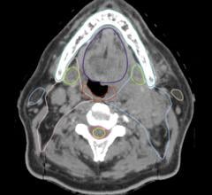
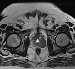
The most difficult challenge in external beam radiation therapy is the ability of doctors to accurately deliver high doses of radiation to a moving tumor without damaging the surrounding healthy tissue.
With organs such as the prostate gland and lung, the patient’s breathing moves the position of the tumor while the radiation beam is on. During treatment, the prostate may drift up to 1 cm for more than two minutes. The surrounding tissue is unavoidably radiated. This can result in negative side effects, including urinary and bowel frequency and fatigue.
Stereotactic body radiation therapy (SBRT) is a radiotherapy technique for treating moving targets. To achieve the most precision with SBRT, the linear accelerator relies on technologies that guide the multileaf collimator.
The question now is, what is the best solution to use: robotics, intrafraction-image guidance, gating or 4-D imaging?
SBRT’s Precision Advantage
In SBRT, a specially designed coordinate system is used for the exact localization of the tumors in the body in order to treat it with limited but highly precise treatment fields. SBRT involves the delivery of a single high-dose radiation treatment or a few fractionated radiation treatments, usually up to five treatments. A highly potent biological dose of radiation is delivered to the tumor, improving the cure rates for the tumor in a manner previously not achievable by standard conventional radiation therapy.
The three-year survival rate for patients receiving SBRT was 55.8 percent, compared to the 20 to 35 percent two-year to three-year overall survival rate for studies reporting results from conventional radiation therapy for similar patient groups, a recent study found.
Lead investigator, Robert Timmerman, M.D., professor of radiation oncology, Southwestern Medical Center, Dallas, conducted a study using the Elekta Axesse SBRT linear accelerator to treat nearly 60 patients with inoperable early-stage lung cancer. The 2004-2006 Radiation Therapy Oncology Group (RTOG 0236) study was a phase 2 North American trial of patients with nonsmall cell lung tumors where pre-existing medical conditions precluded surgical treatment.
According to Timmerman, the main finding in this study was the high rate of primary tumor control, which is 97.6 percent at three years.
“SBRT as delivered in this trial provided more than double the rate of primary tumor control than previous reports describing conventional radiation therapy. Primary tumor control is essential for curing lung cancer,” he said.
Timmerman and his team used Elekta SBRT solutions. The system is designed to integrate advanced technologies to enable a radiation therapy treatment technique that delivers highly sculpted dose distributions with exceptional precision.
Real-Time Tracking
According to the American Cancer Society, prostate cancer is the leading cancer in men in the United States, with 192,000 new cases diagnosed each year. Prostate is commonly treated with radiation therapy.
When delivering high doses of radiation to a target, it is critical the dose is delivered accurately to minimize radiation exposure to healthy tissue. Different parts of the body exhibit motion patterns that are unique and distinct, so it’s important to be able to compensate for these shifts when delivering high doses of radiation.
The Accuray CyberKnife, which is approved for use throughout the body, accounts for this by using robotics and intrafraction-image guidance that is capable of adaptive delivery.
“The CyberKnife delivers SBRT-type treatments with accuracy of 1.5 mm or better for tumors that move with respiration and less than 0.8 mm for targets such as the prostate,” reported Omar Dawood, M.D., MPH, vice president of clinical development, Accuray.
When a patient is on the table receiving treatment, the system is designed to automatically retarget beam directions based on the motion patterns of each patient. This eliminates the need to manually reposition the patient during each treatment session.
“The CyberKnife System can take X-ray images every 15 seconds during treatment delivery. This imaging frequency is user-defined, but it also adapts automatically during treatment, based on the tumor motion,” Dawood said. “The system will track bony structure for intracranial tumors or tumors close to the spine. It can also track based on tissue contrast for certain lung tumors. Surrogates, such as gold markers, are needed only when the tumor cannot be located using bony structures or tissue contrast.”
The use of 4-D for CyberKnife is different from other systems, Dawood said. “CyberKnife users may use 4-D CT (computed tomography), but it would only be in the planning stage to better delineate a target,” he explained. “Lung tumors, for example, move with respiration. The position of the tumor between images is predicted based on its historical behavior. The tumor location known from the X-ray images is correlated with chest wall movement measured in real time with an optical tracking system. This breathing model is built just before the treatment and constantly updated during the treatment every time an X-ray image is taken.”
This technology allows the patient to breath normally during the treatment while the robot actively compensates for breathing motion.
4-D Localization
Localization technology is used for precise tracking of tumor targets. The Calypso Medical’s GPS for the Body system uses miniaturized implanted devices, beacon electromagnetic transponders, to continuously track the location of tumors for improved accuracy and management of radiation therapy delivery. The technology is designed for body-wide cancers commonly treated with radiation therapy.
The implanted beacon transponders and the Calypso system provide the clinician continuous, real-time monitoring of the prostate and alert the clinician when the target is outside of acceptable boundaries due to organ motion, thereby enabling corrections during treatment delivery. The technology can adjust the patient position if the tumor moves out of the target zone.
Since the radiation beam is more focused on the tumor target, the Calypso 4-D System — approved for use in the prostate — allows clinicians to contour the radiation dose to the prostate and minimize unwanted dose to adjacent healthy tissues.
“Calypso allows us to track the position of the prostate during prolonged, high-dose SBRT treatments and provides the flexibility to stop the treatment if the prostate moves outside the intended field of radiation,” said Constantine Mantz, M.D., at 21st Century Oncology in Cape Coral, Fla.
What is unique about 4-D on Calypso, Mantz said, is that it tracks the position of the prostate continuously.
Mantz led a study conducted at Stanford University1 to measure side effects using the Calypso-guided dynamic multileaf collimator (DMLC) target tracking with intensity modulated arc therapy (IMAT). Dose distributions to moving targets with DMLC tracking were superior to those without tracking.
“We discovered that rectal, urinary and sexual side effects were significantly diminished with the Calypso system. That is a measure of improved accuracy,” Mantz said.
A work in progress, Dynamic Edge Gating Technology is Calypso’s new solution to improving dose delivery. The system will employ Calypso’s real-time target position information to enable and disable the treatment beam in response to motion of the prostate. The beam can be automatically held when the target position goes outside the motion thresholds and automatically re-enabled when the target is within the motion thresholds.
Targeting Lung Tumors
Lung tumors have long been among the most challenging radiation therapy targets because the patient’s breathing causes tumors to move.
While external skin surface markers or implanted markers are used to estimate lung tumor position during the breathing cycle, the physician can only apply the beam during certain points in the patient’s respiration. These strategies require complex, time-consuming planning and delivery and prolong treatment with an inefficient stop-start beam delivery.
Elekta has introduced technology for SBRT for treating lung tumors. The solution is designed to enable doctors to use 4-D image guidance to confirm the tumor’s location during the breathing cycle. This new technology treats the lesion with a continuous radiation beam, increasing therapy accuracy while using less imaging radiation during treatment delivery.
Elekta’s XVI Symmetry provides tools to manage shifts in the relative positions of the tumor and organs-at-risk during the respiratory cycle. XVI Intuity is applied to ensure the position of the tumor is being tracked, and it accounts for the position of nearby healthy critical structures.
XVI Symmetry and XVI Intuity are feature sets of version 4.5 of Elekta’s X-ray Volume Imaging (XVI) package of software solutions for Image Guided Radiation Therapy (IGRT). XVI 4.5 recently received 510(k) clearance and CE marking to enable sales and distribution in the United States and Europe.
The solutions account for baseline shifts and help physicians deliver treatment using reduced margins around the tumor.
XVI Intuity is designed to extend image guidance by enabling critical structure avoidance, which allows doctors to understand the positional relationship between the target and organs-at-risk. This ensures anatomical changes and corrections to set-up errors have not inadvertently put critical structures into the radiation beam’s path.
Combined Motion Management
Managing tumor motion can be achieved by combining several technologies and techniques. For example, a comprehensive solution may include patient positioning, tumor tracking and gating the treatment beam.
The motion management capabilities on Varian Medical Systems’ Trilogy platform uses gated radiotherapy called RapidArc and interfaces with the Calypso System. This allows clinicians to monitor tumors in real time, gate or turn on and off the beam if the tumor moves outside of a predefined area, and make targeting adjustments when necessary.
Gated RapidArc radiotherapy makes it possible to monitor patient breathing and compensate for tumor motion while quickly delivering a dose during a continuous rotation around the patient. This development
enables the use of RapidArc to target lung tumors with greater precision by gating the beam in response to tumor motion.
To speed up delivery of the radiation, Varian recently added the TrueBeam platform to the Clinac iX and Trilogy accelerators. TrueBeam dynamically synchronizes imaging, patient positioning, motion management and treatment delivery.
It can deliver treatments up to 50 percent faster with a dose delivery rate of up to 2,400 monitor units per minute, which is double the maximum output of earlier, industry-leading Varian systems. This offers greater patient comfort by shortening treatments and may improve precision by leaving less time for tumor motion during dose delivery.
TrueBeam can be used for all forms of advanced external-beam radiotherapy, including image-guided radiotherapy and radiosurgery (IGRT and IGRS), intensity-modulated radiotherapy (IMRT), stereotactic body radiotherapy (SBRT) and RapidArc radiotherapy.
Adaptive Treatment
With image-guided modulated therapy (IGRT), what the physician can do with the images is what matters most.
By integrating a CT scanner into TomoTherapy’s Hi·Art (adaptive radiation therapy) system, the clinician can take CT images for each fraction. Using the CTrue solution, the images are acquired helically, using a fan-beam with a conventional CT detector mounted on a ring gantry. This allows the clinician to verify the location of the tumor. After the CT scan is taken, the radiation therapist makes technical adjustments to accurately align the system to the precise position.
Adding to the treatment options is the TomoDirect solution. Developed as a complement to helical TomoTherapy, both modes use the same binary multileaf collimator and CT-style gantry technology, and share a consistent treatment planning and delivery process. The choice of which modality to use for a given case will depend on the nature of the tumor volume and surrounding organs at risk.
TomoDirect allows clinicians to choose several discrete angles as well as the optimal modulation level required for delivery. It is expected to provide significant time savings in both the planning and delivery phases for several clinical scenarios, including whole breast irradiation and palliative treatments.
In addition to the added capabilities offered by TomoDirect, the Hi·Art system’s treatment modes are being expanded to include 3-D conformal delivery, thereby providing a comprehensive range of options for all clinical cases.
Reference:
1. Keall, et al. “Electromagnetic-Guided DMLC Tracking Enables Motion Management for Intensity Modulated Arc Therapy.” Stanford University, Palo Alto, Calif.

 April 16, 2024


